MyCompetitiveExams Logo MyCompetitiveExams
Search 1,140+ Questions
Chapters, formulas, speed tricks
→
⚡ Brahmastra NEET 2027
720 MARKS

Dynamic 180-question full-length simulator mixed dynamically from all 6 years for 2027 & 2026 aspirants.

Launch Brahmastra 2027 Test →
Start Practice →
100% Free · No Login Required
🧬 Zoology ⭐ High Weightage (~2–3 Qs/Year)

NEET Locomotion and Movement Previous Year Questions with Solutions

Complete repository of official NTA NEET questions for Locomotion and Movement from 2020 to 2026. Every question includes 30-second elimination tricks, step-by-step NCERT solutions, option-by-option traps, and official answer keys.

13 Questions 6 Exam Years (2026, 2025, 2024, 2023, 2021, 2020) 11 Easy · 2 Medium · 0 Challenging
NEET 2026 · Q91 Easy
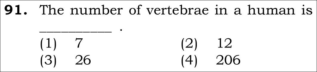
Official NEET 2026 Question 91 - Locomotion and Movement
1
2
3 ✓ Key
4
💡 Verified Solution & 30-Second Hack
✓
Verified Against NTA Final Answer Key
Option (3) : 26
📚 Locomotion and Movement Easy
📋 Given Parameters & Problem Setup
Skeletal Region Adult human vertebral column (backbone)
Vertebral Formula C₇ T₁₂ L₅ S₍₁₎ Co₍₁₎
⚡

The 30-Second Exam Hall Shortcut: ⚡ Adult Vertebral Count Rule

⏱️ Save ~40 seconds

7 Cervical + 12 Thoracic + 5 Lumbar + 1 Sacrum + 1 Coccyx = 26 Vertebrae!

1 Recall adult vertebral column composition:
2 Cervical = 7, Thoracic = 12, Lumbar = 5, Sacral = 1 (fused from 5), Coccygeal = 1 (fused from 4).
3 Sum: 7 + 12 + 5 + 1 + 1 = 26.
4 Look at Option (3): 26. Pick Option (3) in 5 seconds!
🛡️
Why You Can 100% Trust This Shortcut: NCERT Human Physiology Locomotion verbatim: Our vertebral column is formed by 26 serially arranged units called vertebrae and is dorsally placed.
📐

Step-by-Step Rigorous Workflow & Mathematical Proof

Step 1

Anatomy of the Human Axial Skeleton

The vertebral column forms the central axis of the human body, extending from the base of the skull and protecting the spinal cord. In an adult human, it consists of 26 serially arranged articulated units called vertebrae separated by intervertebral discs.

Total Adult Vertebrae=7+12+5+1+1=26\text{Total Adult Vertebrae} = 7 + 12 + 5 + 1 + 1 = 26
  • • Cervical vertebrae: 7 (in neck, including Atlas C1 and Axis C2).
  • • Thoracic vertebrae: 12 (articulate with ribs).
  • • Lumbar vertebrae: 5 (in lower back).
  • • Sacrum: 1 (formed by fusion of 5 sacral vertebrae).
  • • Coccyx: 1 (tail bone, formed by fusion of 4 coccygeal vertebrae).
Step 2

Distinguish Adult vs Embryonic Count

In human infants, there are 33 individual bones. However, upon skeletal maturation, the 5 sacral vertebrae fuse into 1 sacrum, and the 4 coccygeal vertebrae fuse into 1 coccyx. Thus, the adult anatomical standard taught in the core curriculum is 26.

33 embryonic→fusion of sacrum and coccyx26 adult vertebrae33\text{ embryonic} \xrightarrow{\text{fusion of sacrum and coccyx}} 26\text{ adult vertebrae}
  • • Adult functional bones = 26.
Step 3

Conclusion

The total number of vertebrae in a human is 26.

Option (3) =26\text{Option (3) } = 26
  • • Matches Option (3).
⭐

360° Option Elimination Engine Key Feature

Diagnose every choice: Know exactly why the right option works and why distractors fail.

4-Option Deep Scan
1 A
✗ Incorrect
Verdict & Scientific Reason:

Incorrect. 7 is only the number of cervical (neck) vertebrae, not the entire vertebral column.

⚠️
Student Fallacy (Why candidates pick this trap): Mistaking cervical vertebrae count for total vertebrae.
2 B
✗ Incorrect
Verdict & Scientific Reason:

Incorrect. 12 is the number of thoracic vertebrae (or pairs of ribs), not total vertebrae.

⚠️
Student Fallacy (Why candidates pick this trap): Confusing thoracic vertebrae with the complete backbone.
3 C
✓ 100% Correct
Verdict & Scientific Reason:

Exact canonical count: 26 serially arranged vertebrae in adult human skeleton.

💡
Why this is airtight: None; precise anatomical recall.
4 D
✗ Incorrect
Verdict & Scientific Reason:

Incorrect. 206 is the total number of bones in the ENTIRE adult human skeleton.

⚠️
Student Fallacy (Why candidates pick this trap): Confusing total skeletal bones with vertebral count.
⚠️

NTA Trap Alert: Confusing Total Skeleton Bones (206) with Vertebrae (26)

Under exam pressure, students see "bones in human" and immediately rush to select 206 (Option 4). The question specifically asks for VERTEBRAE, which is 26!

🛡️ How to avoid the -1 mark: Read carefully: Total skeleton = 206 bones; Vertebral column = 26 vertebrae.
📖

Core NEET Syllabus Standard

NEET Biology Core Syllabus · Locomotion and Movement Standard Benchmark
Core Scientific Principle Tested:

"The human vertebral column consists of 26 articulated vertebrae: C₇ T₁₂ L₅ S₁ Co₁."
Syllabus Focus: Skeletal system - Axial skeleton and vertebral column
✓ Core NEET UG Syllabus Anchor Syllabus & Academic Standards ↗
Breakfast at 7 (Cervical), Lunch at 12 (Thoracic), Dinner at 5 (Lumbar) + 1 Sacrum + 1 Coccyx = 26!
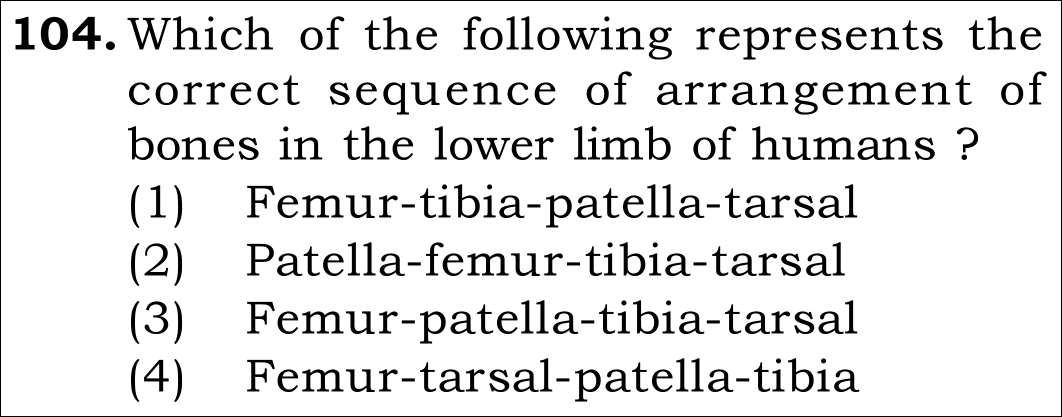
NEET 2026 · Q104 Easy
Official NEET 2026 Question 104 - Locomotion and Movement
1
2
3 ✓ Key
4
💡 Verified Solution & 30-Second Hack
✓
Verified Against NTA Final Answer Key
Option (3) : Femur-patella-tibia-tarsal
📚 Locomotion and Movement Easy
📋 Given Parameters & Problem Setup
Region Human lower limb (leg skeleton)
Required Arrangement Correct anatomical sequence from proximal to distal
⚡

The 30-Second Exam Hall Shortcut: ⚡ Thigh to Ankle Anatomical Ladder

⏱️ Save ~45 seconds

Thigh (Femur) -> Knee cap (Patella) -> Shin (Tibia) -> Ankle (Tarsals)!

1 Start at the hip joint: FEMUR (thigh bone, most proximal).
2 Move down to the knee joint: PATELLA (sesamoid knee cap covering the joint).
3 Move down to the shank: TIBIA (medial weight-bearing shin bone) & Fibula.
4 Move down to the ankle: TARSALS (7 ankle bones).
5 Sequence: Femur -> Patella -> Tibia -> Tarsal.
6 Look at Option (3): Femur-patella-tibia-tarsal. Pick Option (3) in 10 seconds!
🛡️
Why You Can 100% Trust This Shortcut: Standard anatomical sequence of the 30 bones of the human lower limb: Femur (1), Patella (1), Tibia (1), Fibula (1), Tarsals (7), Metatarsals (5), Phalanges (14).
📐

Step-by-Step Rigorous Workflow & Mathematical Proof

Step 1

Review the Bones of the Human Lower Limb

Each human lower limb is made of 30 bones arranged in a linear anatomical hierarchy from proximal (hip articulation) to distal (toes): 1. Femur: Longest, heaviest bone (thigh bone). 2. Patella: Cup-shaped sesamoid bone covering the knee ventrally. 3. Tibia and Fibula: Two parallel long bones of the shank (tibia is medial and larger). 4. Tarsals: Ankle bones (7 in number). 5. Metatarsals: Sole bones (5 in number). 6. Phalanges: Toe bones (14 in number, formula 2,3,3,3,3).

Proximal ⟶ Distal: Femur →Patella →Tibia →Tarsals\text{Proximal } \longrightarrow \text{ Distal: Femur } \to \text{Patella } \to \text{Tibia } \to \text{Tarsals}
  • • Femur articulates with acetabulum proximally and tibia/patella distally.
Step 2

Evaluate the Options Against the Anatomical Sequence

1. Option (1): Femur - tibia - patella - tarsal (Incorrect: places patella distal to tibia). 2. Option (2): Patella - femur - tibia - tarsal (Incorrect: places patella proximal to femur). 3. Option (3): Femur - patella - tibia - tarsal (Correct: strictly proximal to distal order). 4. Option (4): Femur - tarsal - patella - tibia (Incorrect: places tarsals before knee and shin).

Sequence: Femur ⟶ Patella ⟶ Tibia ⟶ Tarsal\text{Sequence: Femur } \longrightarrow \text{ Patella } \longrightarrow \text{ Tibia } \longrightarrow \text{ Tarsal}
  • • Option (3) is the only correct sequence.
Step 3

Conclusion

The correct sequence is Femur-patella-tibia-tarsal.

Matches Option (3)\text{Matches Option (3)}
  • • Matches Option (3).
⭐

360° Option Elimination Engine Key Feature

Diagnose every choice: Know exactly why the right option works and why distractors fail.

4-Option Deep Scan
1 A
✗ Incorrect
Verdict & Scientific Reason:

Incorrect. Patella is situated at the knee between femur and tibia, not below the tibia.

⚠️
Student Fallacy (Why candidates pick this trap): Forgetting the anatomical position of the knee cap.
2 B
✗ Incorrect
Verdict & Scientific Reason:

Incorrect. Femur is the uppermost thigh bone, superior to the patella.

⚠️
Student Fallacy (Why candidates pick this trap): Placing the knee cap above the thigh bone.
3 C
✓ 100% Correct
Verdict & Scientific Reason:

Exact anatomical sequence from top to bottom: Thigh (Femur) -> Knee (Patella) -> Shin (Tibia) -> Ankle (Tarsal).

💡
Why this is airtight: None; flawless appendicular skeletal knowledge.
4 D
✗ Incorrect
Verdict & Scientific Reason:

Incorrect. Tarsals are in the foot/ankle and cannot precede the shin or knee.

⚠️
Student Fallacy (Why candidates pick this trap): Scrambling proximal and distal bone positions.
⚠️

NTA Trap Alert: Confusing Tarsals (Ankle) with Carpals (Wrist)

Carpals belong to the upper limb (wrist); Tarsals belong to the lower limb (ankle). Both are distal to the long bones of the limb.

🛡️ How to avoid the -1 mark: Remember: Legs walk on Tarsals; Hands write with Carpals.
📖

Core NEET Syllabus Standard

NEET Biology Core Syllabus · Locomotion and Movement Standard Benchmark
Core Scientific Principle Tested:

"The human lower limb bones from proximal to distal are Femur, Patella, Tibia/Fibula, Tarsals, Metatarsals, Phalanges."

Syllabus Focus: Appendicular skeleton - Bones of the lower limb
✓ Core NEET UG Syllabus Anchor Syllabus & Academic Standards ↗
From Hip to Toe: Femur first, Patella in the bend, Tibia on the shin, Tarsals at the end!
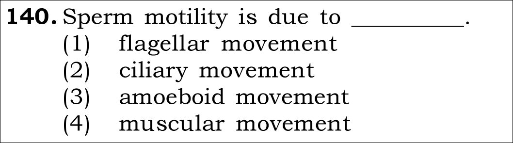
NEET 2026 · Q140 Easy
Official NEET 2026 Question 140 - Locomotion and Movement
1 flagellar movement ✓ Key
2 ciliary movement
3 amoeboid movement
4 muscular movement
💡 Verified Solution & 30-Second Hack
✓
Verified Against NTA Final Answer Key
Option (1) : flagellar movement
📚 Locomotion and Movement Easy
📋 Given Parameters & Problem Setup
Cell Type Human spermatozoon (sperm cell)
Tested Mechanism Type of cellular motility powering forward swimming
⚡

The 30-Second Exam Hall Shortcut: ⚡ Sperm Tail Motility Match

⏱️ Save ~40 seconds

Sperm tail is a Flagellum -> Motility is FLAGELLAR movement!

1 Recall human cell motility types from NCERT Locomotion:
2 Amoeboid movement: Macrophages and leukocytes.
3 Ciliary movement: Ciliated epithelium of respiratory tract and Fallopian tubes.
4 Muscular movement: Skeletal, visceral, and cardiac muscles.
5 Flagellar movement: Spermatozoa swimming via their axial filament/tail!
6 Scan options: Option (1) is flagellar movement.
7 Mark Option (1) in 3 seconds!
🛡️
Why You Can 100% Trust This Shortcut: NCERT Biology · Locomotion and Movement (Amoeboid, Ciliary, and Flagellar Movement in Humans).
📐

Step-by-Step Rigorous Workflow & Mathematical Proof

Step 1

Review Types of Movements Exhibited by Human Cells

Cells of the human body exhibit three main types of movements: amoeboid, ciliary, and muscular, along with flagellar movement exhibited by specialized cells.

Human Cell Movements={Amoeboid:Macrophages, Neutrophils (pseudopodia)Ciliary:Trachea, Oviducts (coordinated cilia)Muscular:Limbs, Jaws, Tongue (myofibrils)Flagellar:Spermatozoa (flagellated tail)\text{Human Cell Movements} = \begin{cases} \text{Amoeboid} &: \text{Macrophages, Neutrophils (pseudopodia)} \\ \text{Ciliary} &: \text{Trachea, Oviducts (coordinated cilia)} \\ \text{Muscular} &: \text{Limbs, Jaws, Tongue (myofibrils)} \\ \mathbf{Flagellar} &: \mathbf{Spermatozoa\text{ (flagellated tail)}} \end{cases}
  • • NCERT explicitly singles out spermatozoa for flagellar movement.
Step 2

Structural Basis of Sperm Flagellum

The sperm tail consists of an axoneme comprising a classical "9+2" microtubule doublet arrangement powered by dynein motor arms. ATP generated by the mitochondrial spiral of the middle piece drives sinusoidal wave propagation along the flagellum, propelling the sperm forward through the female genital tract.

Mitochondrial ATP→Dynein ATPaseFlagellar wave propagation  ⟹  Sperm Swimming\text{Mitochondrial ATP} \xrightarrow{\text{Dynein ATPase}} \text{Flagellar wave propagation} \implies \text{Sperm Swimming}
  • • Ciliary movement propels the non-motile ovum along the Fallopian tube, not the sperm itself.
Step 3

Evaluate Options

Option (1) flagellar movement is the exact mechanism. Option (2) ciliary movement is in oviducts/trachea. Option (3) amoeboid is in phagocytes. Option (4) muscular is in tissues.

Sperm Motility=Flagellar movement  ⟹  Option (1)\text{Sperm Motility} = \mathbf{Flagellar\text{ movement}} \implies \text{Option (1)}
  • • Matches official NTA final answer key Option A / (1).
⭐

360° Option Elimination Engine Key Feature

Diagnose every choice: Know exactly why the right option works and why distractors fail.

4-Option Deep Scan
1 A
✓ 100% Correct
Verdict & Scientific Reason:

100% accurate. Sperm propulsion is driven by the undulatory beating of its flagellar tail.

💡
Why this is airtight: None; standard direct textbook recall.
2 B
✗ Incorrect
Verdict & Scientific Reason:

Incorrect. Cilia line the Fallopian tubes and trachea, but the sperm itself moves via a single flagellum.

⚠️
Student Fallacy (Why candidates pick this trap): Confusing the ciliary lining of the female oviduct with sperm motility.
3 C
✗ Incorrect
Verdict & Scientific Reason:

Incorrect. Amoeboid movement occurs in macrophages and leukocytes via pseudopodia.

⚠️
Student Fallacy (Why candidates pick this trap): Confusing phagocyte diapedesis with sperm propulsion.
4 D
✗ Incorrect
Verdict & Scientific Reason:

Incorrect. Muscle tissue contracts using actin-myosin cross-bridges, not flagellar axonemes.

⚠️
Student Fallacy (Why candidates pick this trap): Confusing uterine myometrial peristalsis with sperm motility.
⚠️

NTA Trap Alert: Confusing Ciliary Movement of Oviduct with Flagellar Movement of Sperm

The ovum is non-motile and transported by ciliated epithelial cells lining the Fallopian tube, whereas the sperm moves under its own power via its flagellum.

🛡️ How to avoid the -1 mark: Remember: Sperm has a Flagellum (Flagellar); Fallopian tube has Cilia (Ciliary).
📖

Core NEET Syllabus Standard

NEET Biology Core Syllabus · Locomotion and Movement Standard Benchmark
Core Scientific Principle Tested:

"Flagellar movement enables the swimming of spermatozoa toward the ovum, driven by the axoneme of the sperm tail."

Syllabus Focus: Types of Movement
✓ Core NEET UG Syllabus Anchor Syllabus & Academic Standards ↗
Cilia carry the Egg in line, but the Flagellar tail swims the Sperm in time!
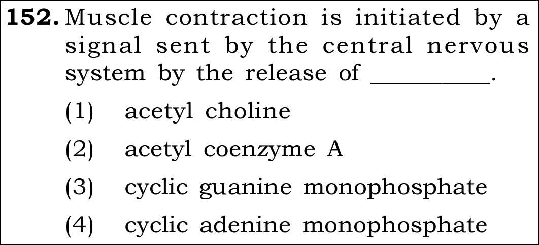
NEET 2026 · Q152 Easy
Official NEET 2026 Question 152 - Locomotion and Movement
1 ✓ Key
2
3
4
💡 Verified Solution & 30-Second Hack
✓
Verified Against NTA Final Answer Key
Option (1) : Acetyl choline
📚 Locomotion and Movement Easy
📋 Given Parameters & Problem Setup
Site of Action Neuromuscular Junction (Motor End Plate)
Trigger CNS motor neuron action potential
Target Effect Action potential generation in sarcolemma
⚡

The 30-Second Exam Hall Shortcut: ⚡ Motor End Plate Gold Standard

⏱️ Save ~40 seconds

Skeletal Neuromuscular Junction = Always Acetylcholine (ACh)!

1 Identify target synapse: Motor neuron junction with skeletal muscle sarcolemma.
2 Recall the primary excitatory neurotransmitter of the vertebrate motor end plate: Acetylcholine.
3 When action potential arrives at axonal terminal, voltage-gated Ca²⁺ influx causes exocytosis of ACh vesicles into the synaptic cleft.
4 ACh binds nicotinic receptors on the sarcolemma, opening Na⁺ channels.
5 Select Option (1): Acetyl choline in 5 seconds.
🛡️
Why You Can 100% Trust This Shortcut: NCERT Biology · Locomotion and Movement (Sliding Filament Theory: Actin filaments slide over myosin during muscle contraction).
📐

Step-by-Step Rigorous Workflow & Mathematical Proof

Step 1

Neuromuscular Junction Organization

A neural signal reaching the neuromuscular junction (the junction between a motor neuron and the sarcolemma of the muscle fibre) releases a neurotransmitter (acetylcholine) which generates an action potential in the sarcolemma.

Axon Terminal Action Potential→Ca2+ InfluxExocytosis of ACh→Synaptic CleftSarcolemma Depolarization\text{Axon Terminal Action Potential} \xrightarrow{\text{Ca}^{2+} \text{ Influx}} \text{Exocytosis of ACh} \xrightarrow{\text{Synaptic Cleft}} \text{Sarcolemma Depolarization}
  • • Vesicles in synaptic knobs fuse with presynaptic membrane and release ACh into synaptic cleft.
Step 2

Excitation-Contraction Coupling

This spreads through the muscle fibre and causes the release of calcium ions into the sarcoplasm from the sarcoplasmic reticulum, initiating sliding filament contraction.

Action Potential→T-tubulesSarcoplasmic Reticulum→Release of Ca2+Troponin C Binding\text{Action Potential} \xrightarrow{\text{T-tubules}} \text{Sarcoplasmic Reticulum} \xrightarrow{\text{Release of Ca}^{2+}} \text{Troponin C Binding}
  • • Acetylcholinesterase subsequently degrades ACh to terminate muscle stimulation.
⭐

360° Option Elimination Engine Key Feature

Diagnose every choice: Know exactly why the right option works and why distractors fail.

4-Option Deep Scan
1 A
✓ 100% Correct
Verdict & Scientific Reason:

100% correct. Acetylcholine is the universal neurotransmitter at vertebrate neuromuscular junctions.

💡
Why this is airtight: None; standard textbook knowledge.
2 B
✗ Incorrect
Verdict & Scientific Reason:

Incorrect. Adrenaline (epinephrine) is an adrenal medullary hormone and sympathetic neurotransmitter, not the motor end plate neurotransmitter.

⚠️
Student Fallacy (Why candidates pick this trap): Confusing sympathetic fight-or-flight hormones with somatic motor neurotransmitters.
3 C
✗ Incorrect
Verdict & Scientific Reason:

Incorrect. Noradrenaline is the postganglionic sympathetic neurotransmitter for smooth muscles and cardiac tissue.

⚠️
Student Fallacy (Why candidates pick this trap): Confusing autonomic nervous system with somatic skeletal motor innervation.
4 D
✗ Incorrect
Verdict & Scientific Reason:

Incorrect. Dopamine is a central nervous system neurotransmitter (involved in reward and basal ganglia motor control), not released at the peripheral neuromuscular junction.

⚠️
Student Fallacy (Why candidates pick this trap): Associating dopamine with motor coordination and wrongly placing it at muscle end plates.
⚠️

NTA Trap Alert: Autonomic vs. Somatic Neurotransmitter Mix-up

Students sometimes confuse sympathetic neurotransmitters (adrenaline/noradrenaline) with somatic motor neuron transmitters. Somatic motor neurons exclusively release acetylcholine onto nicotinic receptors.

🛡️ How to avoid the -1 mark: Remember: Somatic motor neuron -> Skeletal muscle ALWAYS equals Acetylcholine (ACh).
📖

Core NEET Syllabus Standard

NEET Biology Core Syllabus · Locomotion and Movement Standard Benchmark
Core Scientific Principle Tested:

"Neuromuscular junction releases acetylcholine to generate an action potential in the sarcolemma."

Syllabus Focus: Mechanism of Muscle Contraction
✓ Core NEET UG Syllabus Anchor Syllabus & Academic Standards ↗
ACh = Action of Contraction Happens!
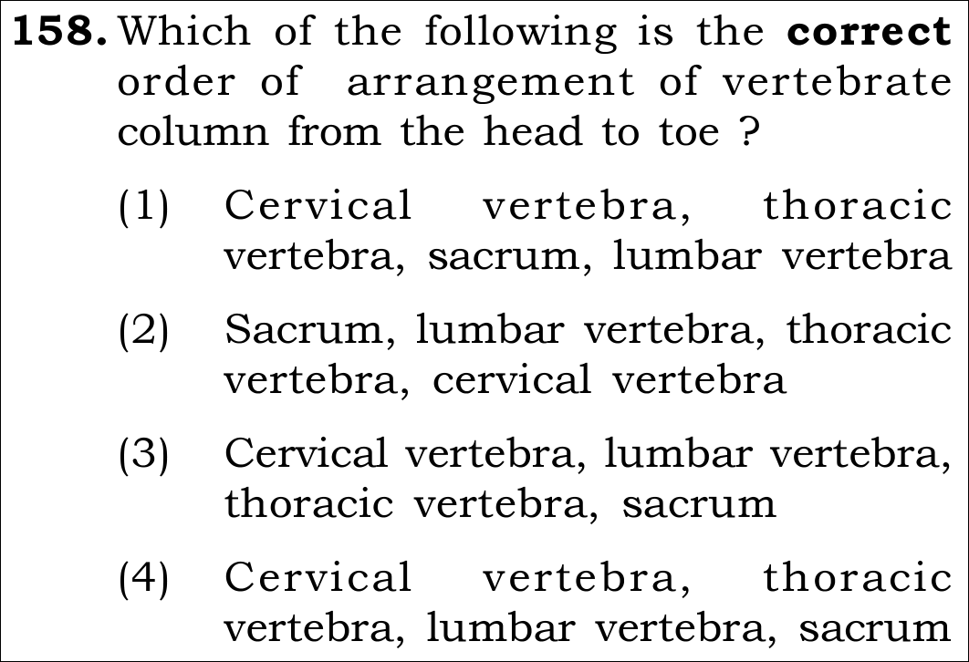
NEET 2026 · Q158 Easy
Official NEET 2026 Question 158 - Locomotion and Movement
1 Cervical vertebra, thoracic vertebra, sacrum, lumbar vertebra
2 Sacrum, lumbar vertebra, thoracic vertebra, cervical vertebra
3 Cervical vertebra, lumbar vertebra, thoracic vertebra, sacrum
4 Cervical vertebra, thoracic vertebra, lumbar vertebra, sacrum ✓ Key
💡 Verified Solution & 30-Second Hack
✓
Verified Against NTA Final Answer Key
Option (4) : (b) -> (a) -> (c) -> (d)
📚 Locomotion and Movement Easy
📋 Given Parameters & Problem Setup
Anatomical Region Human Vertebral Column
Direction of Sequence Cranial (Head) to Caudal (Tail)
Given Parts (a) Thoracic, (b) Cervical, (c) Lumbar, (d) Sacrum
⚡

The 30-Second Exam Hall Shortcut: ⚡ Meal Times Mnemonic (Breakfast at 7, Lunch at 12, Dinner at 5)

⏱️ Save ~30 seconds

Cervical (7) -> Thoracic (12) -> Lumbar (5) -> Sacral (1 fused) -> Coccygeal (1 fused)!

1 Order head to toe: Neck comes first = Cervical (b).
2 Chest comes second = Thoracic (a).
3 Lower back comes third = Lumbar (c).
4 Pelvis comes fourth = Sacrum (d).
5 Target sequence: (b) -> (a) -> (c) -> (d).
6 Select Option (4) in under 5 seconds!
🛡️
Why You Can 100% Trust This Shortcut: NCERT Biology · Locomotion and Movement (Human Skeletal Framework: Axial skeleton consists of 80 bones).
📐

Step-by-Step Rigorous Workflow & Mathematical Proof

Step 1

Vertebral Column Divisions

Our vertebral column is formed by 26 serially arranged units called vertebrae and is dorsally placed. It extends from the base of the skull and constitutes the main framework of the trunk.

Skull Base→Cervical (7)→Thoracic (12)→Lumbar (5)→Sacral (1 fused)→Coccygeal (1 fused)\text{Skull Base} \rightarrow \text{Cervical } (7) \rightarrow \text{Thoracic } (12) \rightarrow \text{Lumbar } (5) \rightarrow \text{Sacral } (1 \text{ fused}) \rightarrow \text{Coccygeal } (1 \text{ fused})
  • • Total 26 vertebrae in adult human skeleton (fused from 33 in embryonic stages).
Step 2

Match Given Labels to Sequence

Align the given anatomical regions: (b) Cervical vertebra -> (a) Thoracic vertebra -> (c) Lumbar vertebra -> (d) Sacrum.

(b)→(a)→(c)→(d)  ⟹  Option (4)(b) \rightarrow (a) \rightarrow (c) \rightarrow (d) \implies \text{Option (4)}
  • • Cranial means superior/headward; caudal means inferior/tailward.
⭐

360° Option Elimination Engine Key Feature

Diagnose every choice: Know exactly why the right option works and why distractors fail.

4-Option Deep Scan
1 A
✗ Incorrect
Verdict & Scientific Reason:

Incorrect. Places lumbar (lower back) before thoracic (chest).

⚠️
Student Fallacy (Why candidates pick this trap): Swapping thoracic and lumbar vertebrae.
2 B
✗ Incorrect
Verdict & Scientific Reason:

Incorrect. Places thoracic before cervical (neck).

⚠️
Student Fallacy (Why candidates pick this trap): Ignoring that cervical vertebrae start directly below the occipital condyles.
3 C
✗ Incorrect
Verdict & Scientific Reason:

Incorrect. Starts with lumbar vertebrae.

⚠️
Student Fallacy (Why candidates pick this trap): Random scrambling.
4 D
✓ 100% Correct
Verdict & Scientific Reason:

100% anatomically accurate. Cervical (b) -> Thoracic (a) -> Lumbar (c) -> Sacrum (d).

💡
Why this is airtight: None; standard anatomical order.
⚠️

NTA Trap Alert: Cranial vs. Caudal Directionality Trap

Ensure you follow Cranial -> Caudal (Top -> Bottom). If a question asks Caudal -> Cranial, the order reverses to Sacrum -> Lumbar -> Thoracic -> Cervical.

🛡️ How to avoid the -1 mark: Read the directional instruction carefully: "cranial (head) to caudal (tail)".
📖

Core NEET Syllabus Standard

NEET Biology Core Syllabus · Locomotion and Movement Standard Benchmark
Core Scientific Principle Tested:

"The vertebral column is differentiated into cervical (7), thoracic (12), lumbar (5), sacral (1-fused) and coccygeal (1-fused) regions starting from the skull."

Syllabus Focus: Human Skeleton · Vertebral Column
✓ Core NEET UG Syllabus Anchor Syllabus & Academic Standards ↗
Can The Lovely Secretary Come? (Cervical, Thoracic, Lumbar, Sacral, Coccygeal)!
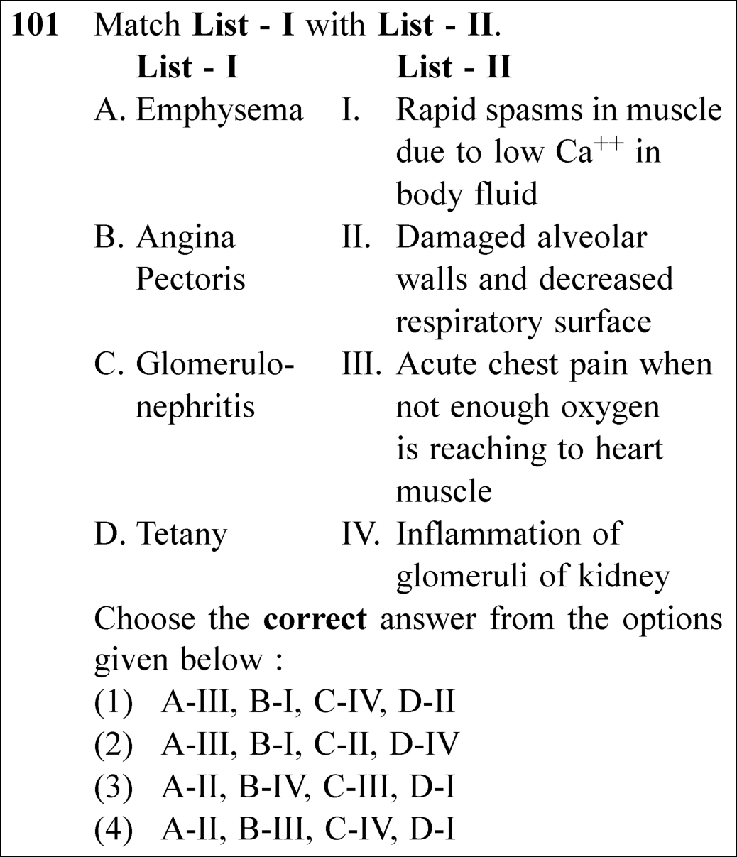
NEET 2025 · Q101 Easy
Official NEET 2025 Question 101 - Locomotion and Movement
1
2
3
4 ✓ Key
💡 Verified Solution & 30-Second Hack
✓
Verified Against NTA Final Answer Key
Option (4) : A-II, B-III, C-IV, D-I
📚 Locomotion and Movement & Human Physiology Easy
📋 Given Parameters & Problem Setup
List I (Disorders) A. Emphysema, B. Angina Pectoris, C. Glomerulonephritis, D. Tetany
List II (Symptoms/Pathology) I. Rapid spasms in muscle due to low Ca⁺⁺ in body fluid, II. Damaged alveolar walls and decreased respiratory surface, III. Acute chest pain when not enough oxygen is reaching to heart muscle, IV. Inflammation of glomeruli of kidney
⚡

The 30-Second Exam Hall Shortcut: ⚡ Physiological Pathology Match

⏱️ Save ~30 seconds

Emphysema = Damaged alveolar walls (A-II). Angina Pectoris = Heart ischemic chest pain (B-III). Glomerulonephritis = Glomerular kidney inflammation (C-IV). Tetany = Low Ca²⁺ muscle spasms (D-I)! ⟹ A-II, B-III, C-IV, D-I!

1 A. Emphysema: Chronic obstructive pulmonary disease where alveolar walls are damaged, reducing surface area for gas exchange ⟹ A matches II.
2 B. Angina Pectoris: Symptom of coronary ischemia characterized by acute retrosternal chest pain due to insufficient oxygen supply to myocardium ⟹ B matches III.
3 C. Glomerulonephritis: Inflammation of the renal glomeruli ⟹ C matches IV.
4 D. Tetany: Hypocalcemic tetany characterized by wild, rapid muscle spasms caused by low extracellular Ca²⁺ ⟹ D matches I.
5 Combined match: A-II, B-III, C-IV, D-I.
6 Select Option (4) in under 10 seconds!
🛡️
Why You Can 100% Trust This Shortcut: NCERT Biology Chapters 17, 18, 19, 20 (Disorders Sections) verbatim.
📐

Step-by-Step Rigorous Workflow & Mathematical Proof

Step 1

Match Emphysema and Angina Pectoris

Examine respiratory and cardiovascular pathology from NCERT:

Emphysema (A)  ⟹  Damaged alveolar walls (II),Angina Pectoris (B)  ⟹  Ischemic chest pain (III)\text{Emphysema (A)} \implies \text{Damaged alveolar walls (II)}, \quad \text{Angina Pectoris (B)} \implies \text{Ischemic chest pain (III)}
  • • A matches II (A-II).
  • • B matches III (B-III).
Step 2

Match Glomerulonephritis and Tetany

Examine excretory and musculoskeletal disorders:

Glomerulonephritis (C)  ⟹  Kidney glomeruli inflammation (IV),Tetany (D)  ⟹  Low Ca2+ muscle spasms (I)\text{Glomerulonephritis (C)} \implies \text{Kidney glomeruli inflammation (IV)}, \quad \text{Tetany (D)} \implies \text{Low Ca}^{2+} \text{ muscle spasms (I)}
  • • C matches IV (C-IV).
  • • D matches I (D-I).
Step 3

Synthesize the Complete Match

Assemble the pairs:

A-II, B-III, C-IV, D-I  ⟹  Option (4)\text{A-II, B-III, C-IV, D-I} \implies \text{Option (4)}
  • • Matches Option (4) perfectly.
⭐

360° Option Elimination Engine Key Feature

Diagnose every choice: Know exactly why the right option works and why distractors fail.

4-Option Deep Scan
1 A
✗ Incorrect
Verdict & Scientific Reason:

Incorrect. Assigns chest pain to emphysema.

⚠️
Student Fallacy (Why candidates pick this trap): Confusing respiratory and cardiac disorders.
2 B
✗ Incorrect
Verdict & Scientific Reason:

Incorrect. Completely scrambled match.

⚠️
Student Fallacy (Why candidates pick this trap): Scrambling multiple organ disorders.
3 C
✗ Incorrect
Verdict & Scientific Reason:

Incorrect. Swaps angina pectoris and glomerulonephritis.

⚠️
Student Fallacy (Why candidates pick this trap): Confusing kidney inflammation with heart pain.
4 D
✓ 100% Correct
Verdict & Scientific Reason:

100% correct. Direct canonical match of all four NCERT disorders.

💡
Why this is airtight: None; flawless medical physiology recall.
⚠️

NTA Trap Alert: Confusing Tetany with Tetanus

Tetanus is caused by Clostridium tetani bacteria (lockjaw). TETANY is caused by low calcium (hypocalcemia) in extracellular fluid, leading to wild muscle twitches and spasms!

🛡️ How to avoid the -1 mark: Remember: Tetany = Low Calcium; Tetanus = Clostridium toxin!
📖

Core NEET Syllabus Standard

NEET Biology Core Syllabus · Locomotion and Movement & Excretory Products Standard Benchmark
Core Scientific Principle Tested:

"Emphysema: damaged alveolar walls; Angina: cardiac chest pain; Glomerulonephritis: inflamed glomeruli; Tetany: low Ca²⁺ spasms."

Syllabus Focus: Disorders of Human Systems (Respiratory, Circulatory, Excretory, Muscular)
✓ Core NEET UG Syllabus Anchor Syllabus & Academic Standards ↗
Emphysema = Alveoli (II), Angina = Chest Pain (III), Nephritis = Glomeruli (IV), Tetany = Low Calcium (I) ⟹ Option 4!
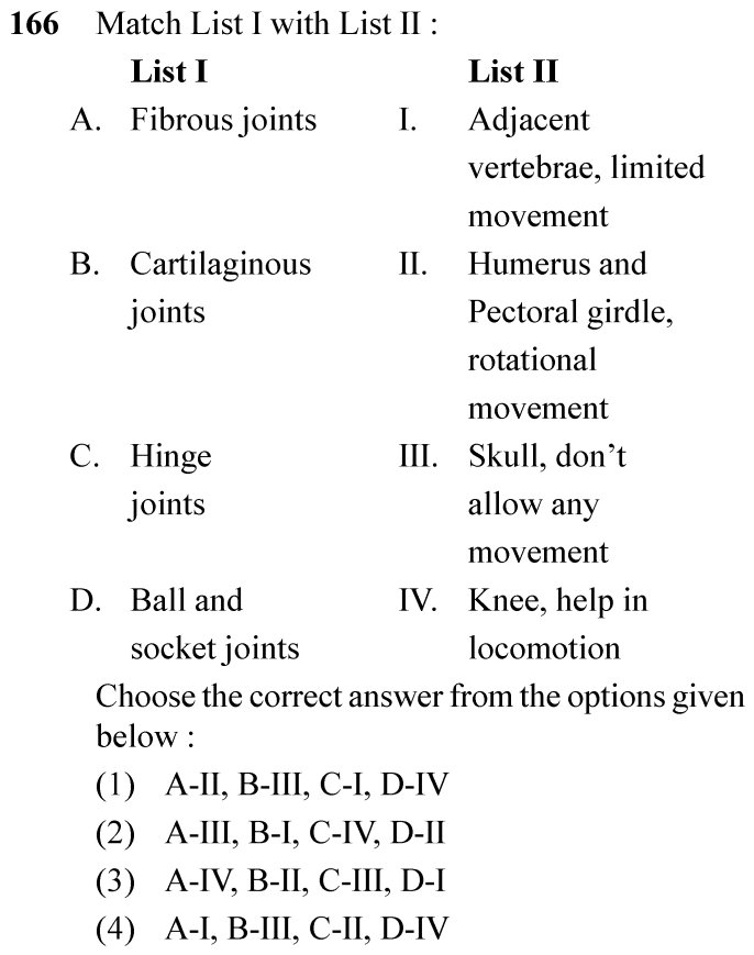
NEET 2024 · Q166 Easy
Official NEET 2024 Question 166 - Locomotion and Movement
1
2 ✓ Key
3
4
💡 Verified Solution & 30-Second Hack
✓
Verified Against NTA Final Answer Key
Option (2) : A-III, B-I, C-IV, D-II
📚 Locomotion and Movement Easy
📋 Given Parameters & Problem Setup
List I (Joint Types) A: Fibrous joints; B: Cartilaginous joints; C: Hinge joints; D: Ball and socket joints
List II (Locations & Movements) I: Adjacent vertebrae, limited movement; II: Humerus and Pectoral girdle, rotational movement; III: Skull, don’t allow any movement; IV: Knee, help in locomotion
⚡

The 30-Second Exam Hall Shortcut: ⚡ Joint Classification Quick Match

⏱️ Save ~25 seconds

Fibrous = Skull sutures (immovable III); Cartilaginous = Intervertebral discs (limited movement I); Hinge = Knee joint (IV); Ball and socket = Shoulder / Humerus & Pectoral girdle (rotational II)! Match: A-III, B-I, C-IV, D-II!

1 A. Fibrous joints: Immovable joints between cranial bones of skull (III).
2 B. Cartilaginous joints: Joint between adjacent vertebrae permit limited movement (I).
3 C. Hinge joints: Synovial joint at knee helping in locomotion (IV).
4 D. Ball and socket joints: Between humerus and glenoid cavity of pectoral girdle permitting multiaxial rotational movement (II).
5 Matching order: A-III, B-I, C-IV, D-II -> Option (2) / B.
🛡️
Why You Can 100% Trust This Shortcut: NCERT Biology · Locomotion and Movement (Classification of Joints)
📐

Step-by-Step Rigorous Workflow & Mathematical Proof

Step 1

Review Immovable and Slightly Movable Joints

Fibrous joints do not allow any movement. This type of joint is shown by the flat skull bones which fuse end-to-end with the help of dense fibrous connective tissues in the form of sutures (A -> III). In cartilaginous joints, the bones involved are joined together with the help of cartilages. The joint between the adjacent vertebrae in the vertebral column is of this pattern and it permits limited movements (B -> I).

Step 2

Review Synovial Joints and Mobility

Hinge joint (knee joint) permits movement primarily in one plane helping in walking/locomotion (C -> IV). Ball and socket joint between humerus and pectoral girdle allows rotational, wide-range movement (D -> II).

Step 3

Select Final Match

A-III, B-I, C-IV, D-II corresponds to Option (2).

⭐

360° Option Elimination Engine Key Feature

Diagnose every choice: Know exactly why the right option works and why distractors fail.

4-Option Deep Scan
1 (1)
✗ Incorrect
Verdict & Scientific Reason:

Mismatches Fibrous joints with Humerus/pectoral girdle.

⚠️
Student Fallacy (Why candidates pick this trap): Mismatches Fibrous joints with Humerus/pectoral girdle.
2 (2)
✓ 100% Correct
Verdict & Scientific Reason:

Accurately pairs all four joint classes with their respective anatomical locations and mechanical mobility.

💡
Why this is airtight: None.
3 (3)
✗ Incorrect
Verdict & Scientific Reason:

Mismatches Fibrous joints with Knee.

⚠️
Student Fallacy (Why candidates pick this trap): Mismatches Fibrous joints with Knee.
4 (4)
✗ Incorrect
Verdict & Scientific Reason:

Mismatches Fibrous joints with vertebrae.

⚠️
Student Fallacy (Why candidates pick this trap): Mismatches Fibrous joints with vertebrae.
⚠️

NTA Trap Alert: Cartilaginous vs Synovial Movement

Thinking intervertebral joints are freely movable synovial joints.

🛡️ How to avoid the -1 mark: Intervertebral discs are cartilaginous (symphysis) allowing only limited, cumulative flexibility.
📖

Core NEET Syllabus Standard

Biology Core Zoology · Locomotion and Movement Standard Benchmark
Core Scientific Principle Tested:

"Fibrous joints do not allow any movement (skull sutures). In cartilaginous joints, bones are joined by cartilage (adjacent vertebrae, limited movement). Synovial joints: Ball and socket joint (between humerus and pectoral girdle), Hinge joint (knee joint)."

Syllabus Focus: Classification of Joints: Fibrous, Cartilaginous, and Synovial Types
✓ Core NEET UG Syllabus Anchor Syllabus & Academic Standards ↗
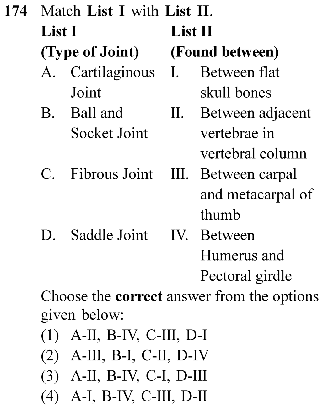
NEET 2023 · Q174 Easy
Official NEET 2023 Question 174 - Locomotion and Movement
1
2
3 ✓ Key
4
💡 Verified Solution & 30-Second Hack
✓
Verified Against NTA Final Answer Key
Option (3) : A-II, B-IV, C-I, D-III
📚 Locomotion and Movement Easy
📋 Given Parameters & Problem Setup
List I (Joint Types) Cartilaginous Joint, Ball and Socket Joint, Fibrous Joint, Saddle Joint
List II (Locations) Between flat skull bones, Between adjacent vertebrae, Between carpal and metacarpal of thumb, Between humerus and pectoral girdle
⚡

The 30-Second Exam Hall Shortcut: ⚡ Skeletal Joint Location Matcher

⏱️ Save ~25 seconds

Match standard NCERT joint examples: Cartilaginous = Between adjacent vertebrae (II). Ball and socket = Between humerus and pectoral girdle (IV). Fibrous = Sutures between flat skull bones (I). Saddle = Between carpal and metacarpal of thumb (III)! Match Option (3)!

1 Cartilaginous joint: Between adjacent vertebrae in vertebral column (permits limited movement) -> A-II.
2 Ball and socket joint: Between head of humerus and glenoid cavity of pectoral girdle -> B-IV.
3 Fibrous joint: Dense fibrous connective tissue forming sutures between flat skull bones (immovable) -> C-I.
4 Saddle joint: Between carpal and metacarpal of thumb -> D-III.
5 Match combination: A-II, B-IV, C-I, D-III -> Option (3).
🛡️
Why You Can 100% Trust This Shortcut: Canonical NCERT Locomotion and Movement anatomical joint classification.
📐

Step-by-Step Rigorous Workflow & Mathematical Proof

Step 1

Anatomical Classification of Joints

Joints are classified into three structural categories based on the tissue joining the articulating bones:

A. Cartilaginous Joint:Intervertebral discs between adjacent vertebrae  ⟹  IIB. Ball and Socket Joint:Synovial joint between humerus and glenoid cavity of pectoral girdle  ⟹  IVC. Fibrous Joint:Immovable sutures between flat bones of cranium  ⟹  ID. Saddle Joint:Synovial joint between carpal and metacarpal of the thumb  ⟹  III\begin{aligned} \text{\textbf{A. Cartilaginous Joint:}} & \quad \text{Intervertebral discs between adjacent vertebrae} \implies \mathbf{\text{II}} \\[4pt] \text{\textbf{B. Ball and Socket Joint:}} & \quad \text{Synovial joint between humerus and glenoid cavity of pectoral girdle} \implies \mathbf{\text{IV}} \\[4pt] \text{\textbf{C. Fibrous Joint:}} & \quad \text{Immovable sutures between flat bones of cranium} \implies \mathbf{\text{I}} \\[4pt] \text{\textbf{D. Saddle Joint:}} & \quad \text{Synovial joint between carpal and metacarpal of the thumb} \implies \mathbf{\text{III}} \end{aligned}
Step 2

Verbatim NCERT Reference

NCERT states under Joints: - "Fibrous joints do not allow any movement... shown by the flat skull bones which fuse end-to-end with the help of dense fibrous connective tissues in the form of sutures." - "In cartilaginous joints, the bones involved are joined together with the help of cartilages... between the adjacent vertebrae in the vertebral column." - "Ball and socket joint (between humerus and pectoral girdle), Saddle joint (between carpal and metacarpal of thumb)."

Step 3

Option Verification

Matching A-II, B-IV, C-I, D-III uniquely corresponds to Option (3).

⭐

360° Option Elimination Engine Key Feature

Diagnose every choice: Know exactly why the right option works and why distractors fail.

4-Option Deep Scan
1 1
✗ Incorrect
Verdict & Scientific Reason:

Mismatches Fibrous joint with thumb and Saddle joint with skull.

⚠️
Student Fallacy (Why candidates pick this trap): Swapping immovable fibrous sutures with movable synovial saddle joints.
2 2
✗ Incorrect
Verdict & Scientific Reason:

Mismatches Cartilaginous joint with thumb and Ball and socket with skull.

⚠️
Student Fallacy (Why candidates pick this trap): Scrambling joint anatomical locations.
3 3
✓ 100% Correct
Verdict & Scientific Reason:

Every joint type matches its canonical NCERT location verbatim.

💡
Why this is airtight: None; textbook verbatim match.
4 4
✗ Incorrect
Verdict & Scientific Reason:

Mismatches Cartilaginous joint with skull bones.

⚠️
Student Fallacy (Why candidates pick this trap): Thinking cranial sutures are cartilaginous.
⚠️

NTA Trap Alert: Hinge vs. Pivot vs. Saddle Synovial Subtype Trap

Students frequently mix up specific synovial joint subtypes: Knee/Elbow = Hinge; Atlas-Axis = Pivot; Carpal-Metacarpal of thumb = Saddle.

🛡️ How to avoid the -1 mark: Remember: Thumb carpal-metacarpal is the classic unique Saddle joint.
📖

Core NEET Syllabus Standard

NCERT Biology · Locomotion and Movement Standard Benchmark
Core Scientific Principle Tested:

"Cartilaginous joints occur between adjacent vertebrae; Ball and socket joint occurs between humerus and pectoral girdle; Fibrous joints occur in skull sutures; Saddle joint occurs between carpal and metacarpal of thumb."

Syllabus Focus: Joints
✓ Core NEET UG Syllabus Anchor Syllabus & Academic Standards ↗
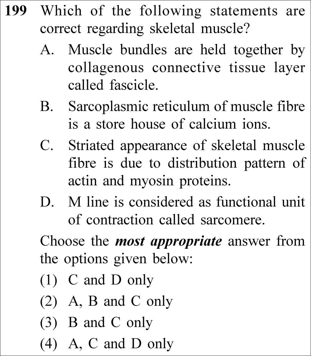
NEET 2023 · Q199 Medium
Official NEET 2023 Question 199 - Locomotion and Movement
1 C and D only
2 A, B and C only
3 B and C only ✓ Key
4 A, C and D only
💡 Verified Solution & 30-Second Hack
✓
Verified Against NTA Final Answer Key
Option (3) : B and C only
📚 Locomotion and Movement Medium
📋 Given Parameters & Problem Setup
Tissue Skeletal muscle fibre
Statements 4 statements (A-D) on fascia/fascicle, sarcoplasmic reticulum, striations, and sarcomere
⚡

The 30-Second Exam Hall Shortcut: ⚡ Fascicle vs. Fascia & Z-line vs. M-line Precision Filter

⏱️ Save ~25 seconds

Muscle bundles (fascicles) are held by connective tissue called FASCIA (not fascicle, so A is false!). The functional unit of contraction (sarcomere) is between two Z LINES (not M line, so D is false!). Statements B (SR is Ca2+ store) and C (striations due to actin/myosin) are TRUE! Match Option (3) B and C only!

1 Statement A: Muscle bundles held by connective tissue layer called fascicle -> False (Bundles ARE fascicles; the connective tissue sheath is FASCIA).
2 Statement B: Sarcoplasmic reticulum is a storehouse of Ca2+ ions -> True.
3 Statement C: Striated appearance is due to distribution pattern of actin and myosin -> True.
4 Statement D: M line is functional unit called sarcomere -> False (Sarcomere is the portion between two successive Z LINES).
5 B and C only -> Select Option (3).
🛡️
Why You Can 100% Trust This Shortcut: Canonical NCERT Locomotion and Movement muscle ultrastructure definitions.
📐

Step-by-Step Rigorous Workflow & Mathematical Proof

Step 1

Evaluation of Statements B and C (Valid Statements)

Analyzing skeletal muscle ultrastructure according to NCERT: - Statement B: The sarcoplasmic reticulum (SRSR) is a specialized endoplasmic reticulum network whose terminal cisternae sequester and release Ca2+Ca^{2+} upon depolarization. (Statement B is TRUE) - Statement C: Myofibrils exhibit alternating isotropic (I) bands and anisotropic (A) bands arising from the parallel interdigitating arrangement of thin actin and thick myosin myofilaments. (Statement C is TRUE)

Statement B:Sarcoplasmic Reticulum≡Storehouse of Ca2+  ⟹  TRUEStatement C:Striations≡Distribution of Actin and Myosin  ⟹  TRUE\begin{aligned} \text{\textbf{Statement B:}} & \quad \text{Sarcoplasmic Reticulum} \equiv \mathbf{\text{Storehouse of }} Ca^{2+} \implies \mathbf{\text{TRUE}} \\[4pt] \text{\textbf{Statement C:}} & \quad \text{Striations} \equiv \mathbf{\text{Distribution of Actin and Myosin}} \implies \mathbf{\text{TRUE}} \end{aligned}
Step 2

Evaluation of Statements A and D (Invalid Statements)

Analyzing the anatomical errors planted in A and D: - Statement A: Each muscle consists of muscle bundles called fascicles. The collagenous connective tissue layer holding these fascicles together is called fascia. Statement A mistakenly calls the connective tissue layer "fascicle". - Statement D: The functional contractile unit of a myofibril is the sarcomere, defined as the portion between two successive Z-lines, NOT the M-line (the M-line is simply a thin fibrous membrane holding thick filaments in the middle of the A-band).

Statement A:Connective tissue sheath=Fascia(≠Fascicle)  ⟹  FALSEStatement D:Sarcomere=Between two successive Z-lines(≠M-line)  ⟹  FALSE\begin{aligned} \text{\textbf{Statement A:}} & \quad \text{Connective tissue sheath} = \mathbf{\text{Fascia}} \quad (\neq \text{Fascicle}) \implies \mathbf{\text{FALSE}} \\[4pt] \text{\textbf{Statement D:}} & \quad \text{Sarcomere} = \mathbf{\text{Between two successive Z-lines}} \quad (\neq \text{M-line}) \implies \mathbf{\text{FALSE}} \end{aligned}
Step 3

Conclusion

Statements B and C are correct, while A and D are false. Option (3) is the required answer.

⭐

360° Option Elimination Engine Key Feature

Diagnose every choice: Know exactly why the right option works and why distractors fail.

4-Option Deep Scan
1 1
✗ Incorrect
Verdict & Scientific Reason:

Includes false statement D (sarcomere is bounded by Z-lines, not M-line).

⚠️
Student Fallacy (Why candidates pick this trap): Confusing the central M-line with the boundary Z-lines.
2 2
✗ Incorrect
Verdict & Scientific Reason:

Includes false statement A (connective tissue sheath is fascia, not fascicle).

⚠️
Student Fallacy (Why candidates pick this trap): Confusing the term fascicle (the bundle) with fascia (the connective tissue sheath).
3 3
✓ 100% Correct
Verdict & Scientific Reason:

Both Statements B (SR is Ca2+ storehouse) and C (striations due to actin and myosin) are verbatim NCERT truths.

💡
Why this is airtight: None; rigorous ultrastructural precision.
4 4
✗ Incorrect
Verdict & Scientific Reason:

Includes false statements A and D.

⚠️
Student Fallacy (Why candidates pick this trap): Accepting the erroneous fascia and M-line substitutions.
⚠️

NTA Trap Alert: Fascicle vs. Fascia Wording Trap

NTA intentionally substituted "fascicle" in place of "fascia" in Statement A. The muscle bundle IS the fascicle; the connective tissue wrapper is the FASCIA.

🛡️ How to avoid the -1 mark: Muscle bundle = Fascicle. Collagenous connective tissue wrapper = Fascia. Sarcomere = Between two Z-lines.
📖

Core NEET Syllabus Standard

NCERT Biology · Locomotion and Movement Standard Benchmark
Core Scientific Principle Tested:

"Sarcoplasmic reticulum is a store house of calcium ions. Striated appearance is due to actin and myosin distribution. Fascicles are held by fascia; Sarcomere is between two Z-lines."

Syllabus Focus: Structure of Contractile Proteins & Muscle Contraction
✓ Core NEET UG Syllabus Anchor Syllabus & Academic Standards ↗
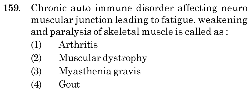
NEET 2021 · Q159 Easy
Official NEET 2021 Question 159 - Locomotion and Movement
1 Arthritis
2 Muscular dystrophy
3 Myasthenia gravis ✓ Key
4 Gout
💡 Verified Solution & 30-Second Hack
✓
Verified Against NTA Final Answer Key
Option (3) : Myasthenia gravis
📚 Locomotion and Movement Easy
📋 Given Parameters & Problem Setup
Condition Traits Chronic autoimmune disorder, affects neuromuscular junction, causes fatigue, muscle weakness, paralysis of skeletal muscle
Candidate Disorders (1) Arthritis, (2) Muscular dystrophy, (3) Myasthenia gravis, (4) Gout
⚡

The 30-Second Exam Hall Shortcut: ⚡ "Autoimmune + Neuromuscular Junction = Myasthenia Gravis"

⏱️ Save ~25 seconds

The verbatim NCERT clinical definition: "Myasthenia gravis: Auto-immune disorder affecting neuromuscular junction leading to fatigue, weakening and paralysis of skeletal muscle." Muscular dystrophy is genetic, arthritis/gout affect joints. Match Option (3) instantly!

1 Key identifier 1: "Autoimmune disorder".
2 Key identifier 2: "Affects neuromuscular junction (NMJ)".
3 Check candidate list: Myasthenia gravis is the only autoimmune disorder of the NMJ.
4 Match Option (3).
🛡️
Why You Can 100% Trust This Shortcut: NCERT Biology · Locomotion and Movement (Disorders of muscular and skeletal system: Myasthenia gravis is autoimmune affecting neuromuscular junction).
📐

Step-by-Step Rigorous Workflow & Mathematical Proof

Step 1

Pathophysiology of Myasthenia Gravis

Myasthenia gravis is a classic autoimmune disease of the peripheral nervous and muscular systems: - Autoantibodies (IgG) target and destroy nicotinic acetylcholine receptors (nAChRs) situated on the sarcolemma of the post-synaptic neuromuscular junction (NMJ). - When acetylcholine is released from motor neurons, fewer receptors are available to generate end-plate potentials. - Over time, repetitive muscular contraction fails, precipitating severe fatigue, flaccid weakness, and eventual paralysis of skeletal muscles (frequently initiating with ptosis of eyelids and diplopia).

Autoantibodies (IgG)→block / destroyACh Receptors at NMJ  ⟹  {↓Neuromuscular transmissionMuscle fatigue & paralysis\text{Autoantibodies (IgG)} \xrightarrow{\text{block / destroy}} \text{ACh Receptors at NMJ} \implies \begin{cases} \downarrow \text{Neuromuscular transmission} \\[2pt] \text{Muscle fatigue \& paralysis} \end{cases}
Step 2

Differential Diagnosis of Listed Disorders

- Arthritis: Inflammation of one or more joints, causing pain and stiffness. - Muscular dystrophy: Progressive degeneration of skeletal muscle caused by an X-linked recessive genetic defect (dystrophin gene mutation), NOT an autoimmune disorder. - Gout: Inflammation of joints caused by accumulation and crystallization of uric acid salts.

⭐

360° Option Elimination Engine Key Feature

Diagnose every choice: Know exactly why the right option works and why distractors fail.

4-Option Deep Scan
1 1
✗ Incorrect
Verdict & Scientific Reason:

Incorrect. Arthritis involves inflammation of joints, not an autoimmune block of neuromuscular junctions.

⚠️
Student Fallacy (Why candidates pick this trap): Rheumatoid arthritis is autoimmune, but it targets synovial membranes in joints, not neuromuscular junctions.
2 2
✗ Incorrect
Verdict & Scientific Reason:

Incorrect. Muscular dystrophy is a genetic defect (often Duchenne muscular dystrophy), not an autoimmune disorder.

⚠️
Student Fallacy (Why candidates pick this trap): The classic pitfall: students remember muscular dystrophy causes progressive muscle weakness, forgetting its genetic etiology.
3 3
✓ 100% Correct
Verdict & Scientific Reason:

Correct. Verbatim matches NCERT definition: chronic autoimmune condition targeting the neuromuscular junction leading to skeletal muscle fatigue and paralysis.

💡
Why this is airtight: None. Accurate diagnostic recognition.
4 4
✗ Incorrect
Verdict & Scientific Reason:

Incorrect. Gout is a metabolic disorder resulting from hyperuricemia and urate crystals in joint spaces.

⚠️
Student Fallacy (Why candidates pick this trap): Confusing joint pathology with neuromuscular junction signaling failure.
⚠️

NTA Trap Alert: Autoimmune (Myasthenia) vs Genetic (Muscular Dystrophy) Distinction

NTA loves contrasting Myasthenia Gravis and Muscular Dystrophy. Both lead to muscle wasting and weakness, but their fundamental causes are entirely different.

🛡️ How to avoid the -1 mark: Anchor: Myasthenia = auto-iMmune (NMJ antibodies); Dystrophy = genetic Defect (dystrophin gene).
📖

Core NEET Syllabus Standard

NEET / NCERT Biology · Locomotion and Movement Standard Benchmark
Core Scientific Principle Tested:

"Myasthenia gravis is an autoimmune disorder affecting neuromuscular junctions, causing skeletal muscle weakness and paralysis."

Syllabus Focus: Disorders of Muscular and Skeletal System
✓ Core NEET UG Syllabus Anchor Syllabus & Academic Standards ↗
NEET 2021 · Q187 Medium
Official NEET 2021 Question 187 - Locomotion and Movement
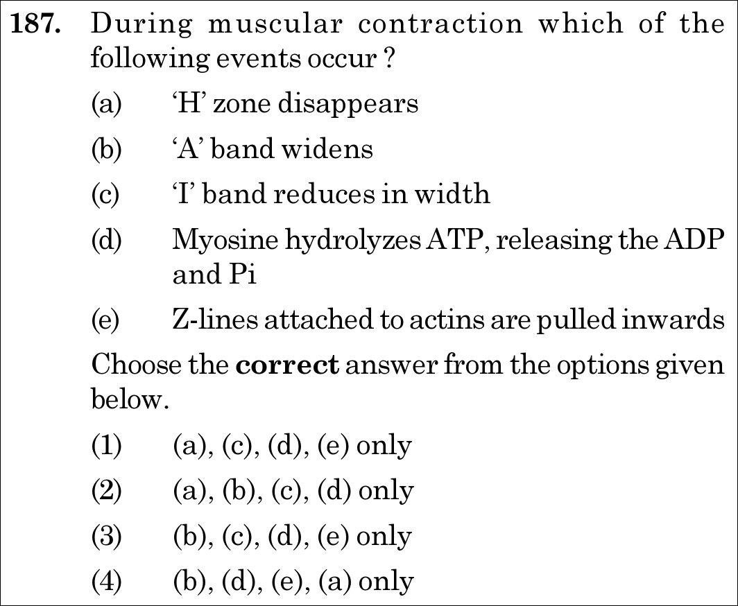
1 (a), (c), (d), (e) only ✓ Key
2 (a), (b), (c), (d) only
3 (b), (c), (d), (e) only
4 (b), (d), (e), (a) only
💡 Verified Solution & 30-Second Hack
✓
Verified Against NTA Final Answer Key
Option (1) : (a), (c), (d), (e) only
📚 Locomotion and Movement Medium
📋 Given Parameters & Problem Setup
Event (a) ‘H’ zone disappears
Event (b) ‘A’ band widens
Event (c) ‘I’ band reduces in width
Event (d) Myosine hydrolyzes ATP, releasing the ADP and Pi
Event (e) Z-lines attached to actins are pulled inwards
⚡

The 30-Second Exam Hall Shortcut: ⚡ "The A-Band NEVER Changes! Eliminate (b) to Win Instantly!"

⏱️ Save ~30 seconds

According to the Sliding Filament Theory, the Anisotropic (A) band length is determined by the length of thick myosin filaments and NEVER widens or changes during contraction. Statement (b) is completely FALSE! Eliminate any option containing (b) -> Options 2, 3, and 4 are destroyed instantly!

1 Universal Law of Muscle Contraction: The A-band retains its length (constant length); it NEVER widens.
2 Inspect options for false statement (b): - Option (2) has (b) -> Eliminate - Option (3) has (b) -> Eliminate - Option (4) has (b) -> Eliminate.
3 Only Option (1) remains: (a), (c), (d), (e) only.
4 Verified in under 10 seconds!
🛡️
Why You Can 100% Trust This Shortcut: NCERT Biology · Locomotion and Movement (Mechanism of Muscle Contraction: During contraction, the ‘I’ bands get reduced, whereas the ‘A’ bands retain the length. The ‘H’ zone disappears as actin filaments overlap).
📐

Step-by-Step Rigorous Workflow & Mathematical Proof

Step 1

Sliding Filament Mechanism (Huxley & Hanson Model)

Muscle contraction occurs via the sliding of thin (actin) filaments over thick (myosin) filaments without any change in the physical length of the individual filaments themselves: - (d) ATP Hydrolysis: The globular myosin head acts as an ATPase enzyme, hydrolyzing ATP into ADP+Pi\text{ADP} + \text{P}_i. This energizes the head into a cocked high-energy state that binds the exposed active sites on actin to form a cross-bridge. - Power Stroke & Z-Line Movement: Release of ADP\text{ADP} and Pi\text{P}_i triggers the power stroke, tilting the myosin head and pulling the attached actin filaments inward toward the centre of the sarcomere (MM-line). Consequently, the Z-lines attached to actins are pulled inwards ((e) is True). - (c) I-Band Shortening: The isotropic (I) bands (composed of actin filaments only) narrow and reduce in width ((c) is True). - (a) Disappearance of H-Zone: The central H-zone (the region of thick myosin not overlapped by thin actin at rest) progressively narrows and disappears as opposing actin filaments overlap ((a) is True). - (b) Constancy of A-Band: The anisotropic (A) band corresponds to the entire physical length of the myosin filament. Because myosin filaments do not lengthen or expand, the A-band retains its constant length and NEVER widens ((b) is FALSE).

Step 2

Sarcomere Dimensional Invariants Summary

Sarcomere MetricStatus During ContractionSarcomere Length (Z to Z)ShortensI-Band (Actin only)Reduces in width (Shortens)H-Zone (Myosin only)Narrows and DisappearsA-Band (Total Myosin)Retains Length (Constant, DOES NOT WIDEN)\begin{array}{l|l} \textbf{Sarcomere Metric} & \textbf{Status During Contraction} \\ \hline \text{Sarcomere Length (Z to Z)} & \text{Shortens} \\ \text{I-Band (Actin only)} & \text{Reduces in width (Shortens)} \\ \text{H-Zone (Myosin only)} & \text{Narrows and Disappears} \\ \mathbf{A\text{-Band (Total Myosin)}} & \mathbf{Retains\,Length\,(Constant,\,DOES\,NOT\,WIDEN)} \\ \end{array} Therefore, the events that occur are (a), (c), (d), and (e) only.

Contraction Events  ⟹  {(a) H-zone disappears, (c) I-band reduces, (d) ATP hydrolyzed, (e) Z-lines pulled inward}\textbf{Contraction Events} \implies \{ \text{(a) } H\text{-zone disappears}, \, \text{(c) } I\text{-band reduces}, \, \text{(d) ATP hydrolyzed}, \, \text{(e) } Z\text{-lines pulled inward} \}
⭐

360° Option Elimination Engine Key Feature

Diagnose every choice: Know exactly why the right option works and why distractors fail.

4-Option Deep Scan
1 1
✓ 100% Correct
Verdict & Scientific Reason:

Correct. Includes all four verified contraction events while correctly excluding the false statement (b).

💡
Why this is airtight: None. Candidate recognizes the constancy of the A-band.
2 2
✗ Incorrect
Verdict & Scientific Reason:

Incorrect because (b) claims the A-band widens, which is physiologically impossible.

⚠️
Student Fallacy (Why candidates pick this trap): Believing that "contraction" means bands widen, or confusing A-band with sarcomere changes.
3 3
✗ Incorrect
Verdict & Scientific Reason:

Incorrect due to inclusion of false statement (b).

⚠️
Student Fallacy (Why candidates pick this trap): Failing to eliminate the A-band widening distractor.
4 4
✗ Incorrect
Verdict & Scientific Reason:

Incorrect due to inclusion of false statement (b).

⚠️
Student Fallacy (Why candidates pick this trap): Overlooking the invariance of the A-band.
⚠️

NTA Trap Alert: The "A-Band Widens" Optical Illusion Trap

When muscles contract, they bulge in macroscopic thickness. Careless candidates confuse gross muscle bulging with the microscopic sarcomere A-band, assuming the A-band must widen. The A-band length is fixed by myosin filament length.

🛡️ How to avoid the -1 mark: Remember: A-band is Anchor: It ALWAYS stays constant.
📖

Core NEET Syllabus Standard

NEET / NCERT Biology · Locomotion and Movement Standard Benchmark
Core Scientific Principle Tested:

"During contraction, H-zone disappears, I-band narrows, Z-lines move inward, and A-band retains constant length."

Syllabus Focus: Mechanism of Muscle Contraction
✓ Core NEET UG Syllabus Anchor Syllabus & Academic Standards ↗
NEET 2021 · Q188 Easy
Official NEET 2021 Question 188 - Locomotion and Movement
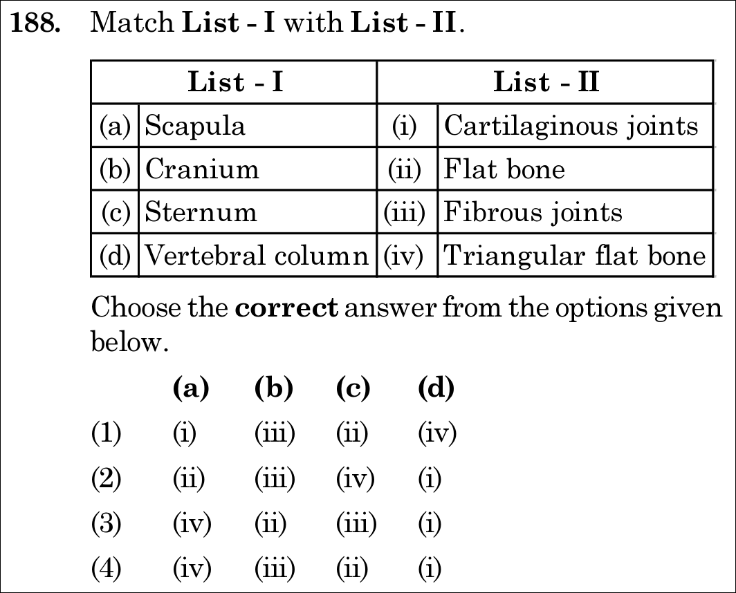
1 (i) (iii) (ii) (iv)
2 (ii) (iii) (iv) (i)
3 (iv) (ii) (iii) (i)
4 (iv) (iii) (ii) (i) ✓ Key
💡 Verified Solution & 30-Second Hack
✓
Verified Against NTA Final Answer Key
Option (4) : (a)-(iv), (b)-(iii), (c)-(ii), (d)-(i)
📚 Locomotion and Movement Easy
📋 Given Parameters & Problem Setup
List I (Bones / Regions) (a) Scapula, (b) Cranium, (c) Sternum, (d) Vertebral column
List II (Characteristics / Joint Types) (i) Cartilaginous joints, (ii) Flat bone, (iii) Fibrous joints, (iv) Triangular flat bone
⚡

The 30-Second Exam Hall Shortcut: ⚡ "Scapula = Triangular Flat Bone, Cranium = Fibrous Sutures"

⏱️ Save ~25 seconds

Scapula is a large triangular flat bone situated on the dorsal thorax ((a)-(iv)), and cranial bones are united by immovable fibrous joints / sutures ((b)-(iii)). Looking at options, only Option (4) matches both (a)-(iv) and (b)-(iii)! Solved in 10 seconds.

1 Match (a) Scapula -> (iv) Triangular flat bone (pectoral girdle).
2 Match (b) Cranium -> (iii) Fibrous joints (dense fibrous connective tissue sutures).
3 Match (c) Sternum -> (ii) Flat bone (ventral midline of thorax).
4 Match (d) Vertebral column -> (i) Cartilaginous joints (intervertebral discs permitting limited movement).
5 Match pattern (iv), (iii), (ii), (i) -> Option (4).
🛡️
Why You Can 100% Trust This Shortcut: NCERT Biology · Locomotion and Movement (Skeletal System & Joints: Scapula is a large triangular flat bone; Cranium bones are fused by fibrous joints; Sternum is a flat bone on the ventral midline; Adjacent vertebrae in vertebral column are joined by cartilaginous joints).
📐

Step-by-Step Rigorous Workflow & Mathematical Proof

Step 1

Morphological and Histological Characterization of Skeletal Elements

- (a) Scapula: A large, thin, triangular flat bone situated in the dorsal part of the thorax between the second and seventh ribs. It features a spine terminating in the acromion process. - (b) Cranium: Cranial bones are firmly knit together end-to-end by dense fibrous connective tissue in the form of sutures to form the brain box; these are classic fibrous joints that permit zero movement (synarthroses). - (c) Sternum: A dagger-shaped flat bone positioned along the ventral midline of the thorax, providing anterior anchorage for true ribs. - (d) Vertebral column: Adjacent vertebrae are articulated by intervertebral fibrocartilaginous discs, which constitute cartilaginous joints (amphiarthroses) permitting limited flexibility and shock absorption.

Scapula  ⟺  Triangular flat bone[(a)→(iv)]Cranium  ⟺  Fibrous joints[(b)→(iii)]Sternum  ⟺  Flat bone[(c)→(ii)]Vertebral column  ⟺  Cartilaginous joints[(d)→(i)]\begin{aligned} \text{Scapula} &\iff \text{Triangular flat bone} \quad [\text{(a)} \to \text{(iv)}] \\ \text{Cranium} &\iff \text{Fibrous joints} \quad [\text{(b)} \to \text{(iii)}] \\ \text{Sternum} &\iff \text{Flat bone} \quad [\text{(c)} \to \text{(ii)}] \\ \text{Vertebral column} &\iff \text{Cartilaginous joints} \quad [\text{(d)} \to \text{(i)}] \end{aligned}
Step 2

Option Alignment

Matching sequence is: (a)-(iv), (b)-(iii), (c)-(ii), (d)-(i). This corresponds perfectly to Option (4).

⭐

360° Option Elimination Engine Key Feature

Diagnose every choice: Know exactly why the right option works and why distractors fail.

4-Option Deep Scan
1 1
✗ Incorrect
Verdict & Scientific Reason:

Incorrect. Links scapula to cartilaginous joints and vertebral column to triangular flat bone.

⚠️
Student Fallacy (Why candidates pick this trap): Swapping Scapula and Vertebral column.
2 2
✗ Incorrect
Verdict & Scientific Reason:

Incorrect. Assigns simple flat bone to scapula and triangular flat bone to sternum (sternum is not triangular).

⚠️
Student Fallacy (Why candidates pick this trap): Failing to remember the triangular contour of the scapula.
3 3
✗ Incorrect
Verdict & Scientific Reason:

Incorrect. Assigns flat bone to cranium and fibrous joints to sternum.

⚠️
Student Fallacy (Why candidates pick this trap): Misaligning cranial sutures.
4 4
✓ 100% Correct
Verdict & Scientific Reason:

Correct. Accurately links all bones and their joint/structural definitions.

💡
Why this is airtight: None. Candidate recalls precise textbook anatomical definitions.
⚠️

NTA Trap Alert: Scapula vs Sternum "Flat Bone" Discrimination Trap

Both scapula and sternum are flat bones. However, NCERT specifically qualifies the scapula as a "triangular flat bone" and sternum simply as a "flat bone on the ventral midline".

🛡️ How to avoid the -1 mark: Identify the shape adjective: "Triangular" belongs uniquely to the Scapula.
📖

Core NEET Syllabus Standard

NEET / NCERT Biology · Locomotion and Movement Standard Benchmark
Core Scientific Principle Tested:

"Scapula is a triangular flat bone; Cranium has fibrous joints; Sternum is a flat bone; Vertebral column has cartilaginous joints."

Syllabus Focus: Skeletal System and Joints
✓ Core NEET UG Syllabus Anchor Syllabus & Academic Standards ↗
NEET 2020 · Q84 Easy
Official NEET 2020 Question 84 - Locomotion and Movement
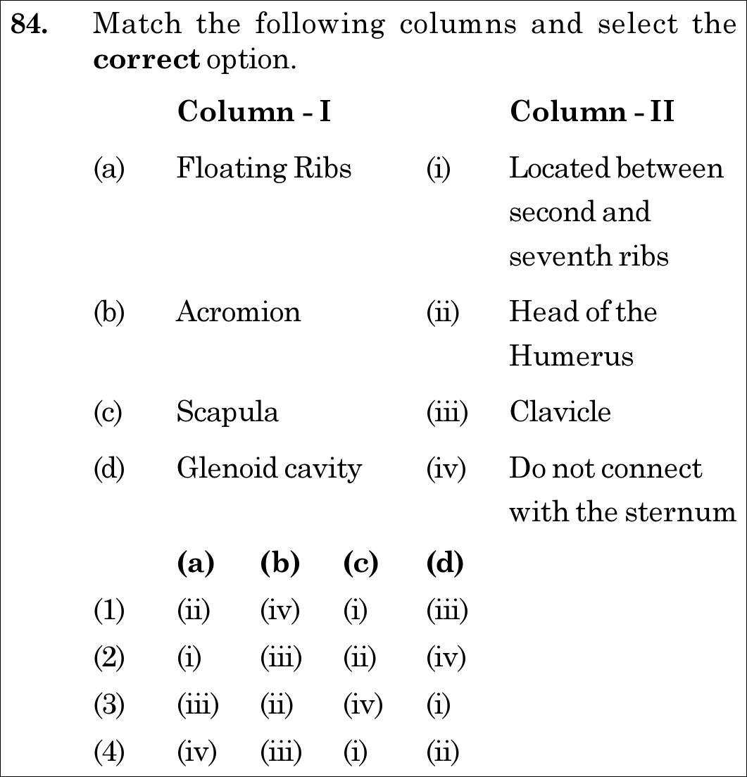
1 (ii) (iv) (i) (iii)
2 (i) (iii) (ii) (iv)
3 (iii) (ii) (iv) (i)
4 (iv) (iii) (i) (ii) ✓ Key
💡 Verified Solution & 30-Second Hack
✓
Verified Against NTA Final Answer Key
Option (4) : (a)-(iv), (b)-(iii), (c)-(i), (d)-(ii)
📚 Locomotion and Movement Easy
📋 Given Parameters & Problem Setup
Column I (Skeletal Elements) (a) Floating Ribs, (b) Acromion, (c) Scapula, (d) Glenoid cavity
Column II (Anatomical Relations) (i) Located between second and seventh ribs, (ii) Head of the Humerus, (iii) Clavicle, (iv) Do not connect with the sternum
⚡

The 30-Second Exam Hall Shortcut: ⚡ "Floating = No Sternum; Acromion = Clavicle; Scapula = 2nd-7th Ribs; Glenoid = Humerus!"

⏱️ 25 seconds

Floating ribs (11th and 12th pairs) do not connect ventrally with the sternum (a->iv). Acromion process articulates with the clavicle (b->iii). Scapula is a large triangular bone situated between 2nd and 7th ribs (c->i). Glenoid cavity articulates with the head of the humerus (d->ii). Combination is (iv), (iii), (i), (ii) -> Option 4 in 10 seconds!

1 Floating ribs (11th & 12th pairs) are not connected ventrally to sternum -> (a)-(iv).
2 Acromion process of scapula articulates with the clavicle (collar bone) -> (b)-(iii).
3 Scapula is situated in dorsal part of thorax between 2nd and 7th ribs -> (c)-(i).
4 Glenoid cavity articulates with head of the humerus forming the ball-and-socket shoulder joint -> (d)-(ii).
5 Mapping: (a)-(iv), (b)-(iii), (c)-(i), (d)-(ii) -> Option 4.
🛡️
Why You Can 100% Trust This Shortcut: NCERT Biology · Human Physiology: Locomotion and Movement (Skeletal System: Ribs & Pectoral Girdle)
📐

Step-by-Step Rigorous Workflow & Mathematical Proof

Step 1

Match Floating Ribs

The last 2 pairs of ribs (11th and 12th) are not connected ventrally to the sternum and are therefore called floating ribs. Thus, (a) matches (iv).

Step 2

Match Acromion

The dorsal, flat, triangular body of the scapula has a slightly elevated ridge called the spine which projects as a flat, expanded process called the acromion. The clavicle articulates with this acromion process. Thus, (b) matches (iii).

Step 3

Match Scapula

Scapula is a large triangular flat bone situated in the dorsal part of the thorax between the second and the seventh ribs. Thus, (c) matches (i).

Step 4

Match Glenoid Cavity

Below the acromion is a shallow depression called the glenoid cavity which articulates with the head of the humerus to form the shoulder joint. Thus, (d) matches (ii).

⭐

360° Option Elimination Engine Key Feature

Diagnose every choice: Know exactly why the right option works and why distractors fail.

4-Option Deep Scan
1 A
✗ Incorrect
Verdict & Scientific Reason:

Incorrect; pairs floating ribs with head of humerus and acromion with sternum disconnection.

⚠️
Student Fallacy (Why candidates pick this trap): Anatomical misalignment.
2 B
✗ Incorrect
Verdict & Scientific Reason:

Incorrect; places floating ribs between second and seventh ribs.

⚠️
Student Fallacy (Why candidates pick this trap): Confusing rib cage boundary markers with floating ribs.
3 C
✗ Incorrect
Verdict & Scientific Reason:

Incorrect; attributes clavicle articulation to floating ribs.

⚠️
Student Fallacy (Why candidates pick this trap): Arbitrary matching.
4 D
✓ 100% Correct
Verdict & Scientific Reason:

Accurate pairing across all four skeletal structures of the human thorax and pectoral girdle.

💡
Why this is airtight: None; textbook osteology definitions.
⚠️

NTA Trap Alert: Scapula Rib Landmarks

Scapula lies dorsally between the 2nd and 7th ribs. Floating ribs are the 11th and 12th ribs (not connected to sternum).

🛡️ How to avoid the -1 mark: Remember: Scapula = 2nd to 7th ribs; Glenoid = Head of humerus; Acromion = Clavicle!
📖

Core NEET Syllabus Standard

Zoology · Locomotion and Movement Standard Benchmark
Core Scientific Principle Tested:

""

Syllabus Focus: Anatomy of Ribs and Pectoral Girdle: Floating Ribs, Acromion, Scapula & Glenoid Cavity
✓ Core NEET UG Syllabus Anchor Syllabus & Academic Standards ↗

Solve 4 NEET Questions a Day — One per Subject

Cover all 1,140 official NEET questions in 285 days at your own pace. Progress is saved on your device. No login needed.