MyCompetitiveExams Logo MyCompetitiveExams
Search 1,140+ Questions
Chapters, formulas, speed tricks
→
⚡ Brahmastra NEET 2027
720 MARKS

Dynamic 180-question full-length simulator mixed dynamically from all 6 years for 2027 & 2026 aspirants.

Launch Brahmastra 2027 Test →
Start Practice →
100% Free · No Login Required
🌿 Botany ⭐ High Weightage (~2–3 Qs/Year)

NEET Anatomy of Flowering Plants Previous Year Questions with Solutions

Complete repository of official NTA NEET questions for Anatomy of Flowering Plants from 2020 to 2026. Every question includes 30-second elimination tricks, step-by-step NCERT solutions, option-by-option traps, and official answer keys.

12 Questions 5 Exam Years (2025, 2024, 2023, 2021, 2020) 9 Easy · 3 Medium · 0 Challenging
NEET 2025 · Q118 Easy
Official NEET 2025 Question 118 - Anatomy of Flowering Plants
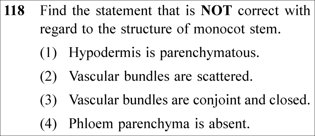
1 Hypodermis is parenchymatous. ✓ Key
2 Vascular bundles are scattered.
3 Vascular bundles are conjoint and closed.
4 Phloem parenchyma is absent.
💡 Verified Solution & 30-Second Hack
✓
Verified Against NTA Final Answer Key
Option (1) : Hypodermis is parenchymatous.
📚 Anatomy of Flowering Plants Easy
📋 Given Parameters & Problem Setup
Plant Organ Monocotyledonous stem (e.g., Zea mays / maize stem)
Target Statement Statement that is NOT correct (False statement)
⚡

The 30-Second Exam Hall Shortcut: ⚡ Monocot vs Dicot Stem Hypodermis Fingerprint

⏱️ Save ~25 seconds

Dicot Stem = Collenchymatous hypodermis! Monocot Stem = SCLERENCHYMATOUS hypodermis! Statement (1) claiming parenchymatous hypodermis is completely WRONG!

1 Check each statement against NCERT monocot stem anatomy:
2 Hypodermis is parenchymatous -> FALSE! NCERT verbatim: "The monocotyledonous stem has a sclerenchymatous hypodermis..."
3 Vascular bundles are scattered -> TRUE! Numerous vascular bundles scattered in ground tissue.
4 Vascular bundles are conjoint and closed -> TRUE! Xylem and phloem on same radius, cambium absent (closed).
5 Phloem parenchyma is absent -> TRUE! Monocot vascular bundles lack phloem parenchyma and have water-containing cavities.
6 Statement (1) is NOT correct!
7 Select Option (1)!
🛡️
Why You Can 100% Trust This Shortcut: Dicot vs Monocot Stomatal & Trichome Anatomy (NCERT Biology · Anatomy of Flowering Plants).
📐

Step-by-Step Rigorous Workflow & Mathematical Proof

Step 1

Diagnostic Features of Monocotyledonous Stem Anatomy

Transverse section of a typical monocot stem (e.g., maize, grass) reveals the following distinct anatomical features:

  • • Epidermis: Single outermost layer with cuticle; trichomes (epidermal hairs) are generally absent.
  • • Hypodermis: A few layers of thick-walled, lignified sclerenchymatous cells providing mechanical strength to withstand bending and wind stress. It is NEVER parenchymatous or collenchymatous.
  • • Ground tissue: Large, conspicuous parenchymatous tissue extending from hypodermis to the center without differentiation into cortex, endodermis, pericycle, and pith.
Step 2

Vascular Architecture in Monocot Stem

Vascular bundles display iconic diagnostic hallmarks:

  • • Scattered distribution: A large number of vascular bundles are scattered throughout the ground tissue; peripheral bundles are smaller and more densely arranged than larger central ones.
  • • Bundle sheath: Each vascular bundle is enveloped by a sclerenchymatous bundle sheath.
  • • Conjoint and closed: Vascular bundles are conjoint (xylem and phloem on same radius), collateral, and closed (cambium is absent, precluding secondary growth).
  • • Phloem composition: Composed of sieve tubes and companion cells; phloem parenchyma is absent.
  • • Water cavity: A lysigenous cavity (water-containing cavity) is formed by the breakdown of inner protoxylem vessels.
Step 3

Evaluation of the Options

Statement (1) claims "Hypodermis is parenchymatous", directly contradicting the presence of a sclerenchymatous hypodermis. Hence, Statement (1) is incorrect.

Monocot Hypodermis=Sclerenchymatous≠Parenchymatous\text{Monocot Hypodermis} = \text{Sclerenchymatous} \neq \text{Parenchymatous}
⭐

360° Option Elimination Engine Key Feature

Diagnose every choice: Know exactly why the right option works and why distractors fail.

4-Option Deep Scan
1 A
✓ 100% Correct
Verdict & Scientific Reason:

100% correct choice (as the incorrect statement). Monocot stem hypodermis is composed of sclerenchyma, providing mechanical rigidity.

💡
Why this is airtight: None; knows that hypodermis in monocot stem is sclerenchyma.
2 B
✗ Incorrect
Verdict & Scientific Reason:

Incorrect choice (true statement). Monocot vascular bundles are characteristically scattered throughout parenchymatous ground tissue (atactostele).

⚠️
Student Fallacy (Why candidates pick this trap): Mistaking a correct biological fact as the false statement.
3 C
✗ Incorrect
Verdict & Scientific Reason:

Incorrect choice (true statement). Cambium is absent, making them closed.

⚠️
Student Fallacy (Why candidates pick this trap): Confusing open dicot bundles with closed monocot bundles.
4 D
✗ Incorrect
Verdict & Scientific Reason:

Incorrect choice (true statement). Absence of phloem parenchyma is a textbook hallmark of monocots.

⚠️
Student Fallacy (Why candidates pick this trap): Forgetting that monocots generally lack phloem parenchyma.
⚠️

NTA Trap Alert: Dicot vs Monocot Stem Hypodermis Tissue Type

Students frequently confuse hypodermal tissues: Dicot stem hypodermis is COLLENCHYMATOUS (flexible mechanical support). Monocot stem hypodermis is SCLERENCHYMATOUS (rigid mechanical support). Parenchyma forms the ground tissue, NOT the hypodermis!

🛡️ How to avoid the -1 mark: Dicot = Collenchyma; Monocot = Sclerenchyma; Neither has parenchymatous hypodermis!
📖

Core NEET Syllabus Standard

NEET Biology Core Syllabus · Anatomy of Flowering Plants Standard Benchmark
Core Scientific Principle Tested:

"Monocot stem possesses a sclerenchymatous hypodermis, scattered conjoint closed vascular bundles, and lacks phloem parenchyma."
Syllabus Focus: Monocotyledonous Stem
✓ Core NEET UG Syllabus Anchor Syllabus & Academic Standards ↗
Mo-Scle: Monocot = Sclerenchymatous hypodermis; Di-Col: Dicot = Collenchymatous!
NEET 2024 · Q101 Easy
Official NEET 2024 Question 101 - Anatomy of Flowering Plants
1
2
3 ✓ Key
4
💡 Verified Solution & 30-Second Hack
✓
Verified Against NTA Final Answer Key
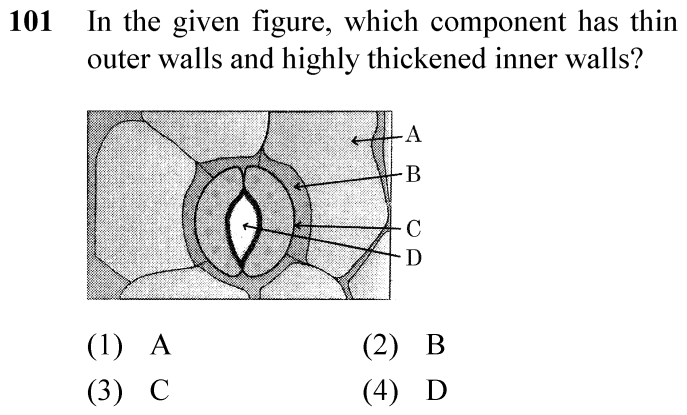
Option (3) : Guard cell
📚 Anatomy of Flowering Plants Easy
📋 Given Parameters & Problem Setup
Tissue System Epidermal Tissue System
Structural Feature Thin outer walls and highly thickened inner walls bordering stomatal aperture
⚡

The 30-Second Exam Hall Shortcut: ⚡ Stomatal Wall Asymmetry Rule

⏱️ Save ~20 seconds

Inner wall facing aperture is thick and inelastic; outer wall is thin and elastic. This asymmetric structure belongs uniquely to Guard Cells!

1 Recall stomatal anatomy:
2 - Subsidiary cells: specialized epidermal cells surrounding guard cells, thin-walled.
3 - Epidermal cells: outer protective layer.
4 - Guard cells: kidney/bean-shaped (or dumb-bell in grasses) with thin outer walls and differentially thickened inner walls facing the pore.
5 Differential thickening is essential for opening during turgidity.
6 Hence, the component is the Guard cell -> Option (3) / C.
🛡️
Why You Can 100% Trust This Shortcut: NCERT Biology · Anatomy of Flowering Plants (Epidermal Tissue System and Stomata)
📐

Step-by-Step Rigorous Workflow & Mathematical Proof

Step 1

Understand Stomatal Apparatus Components

The stomatal apparatus consists of the stomatal aperture (pore), two guard cells flanking the pore, and surrounding specialized subsidiary cells derived from epidermal tissue.

Step 2

Analyze Guard Cell Wall Specialization

In dicots, guard cells are bean-shaped or kidney-shaped. The outer walls of guard cells (away from the stomatal pore) are thin, while the inner walls (facing and bordering the stomatal pore) are highly thickened and inelastic. Cellulose microfibrils are oriented radially rather than longitudinally, facilitating aperture opening when guard cells become turgid.

Step 3

Correlate with Given Question

The question asks which component possesses thin outer walls and highly thickened inner walls. This is the hallmark anatomical definition of guard cells.

⭐

360° Option Elimination Engine Key Feature

Diagnose every choice: Know exactly why the right option works and why distractors fail.

4-Option Deep Scan
1 (1)
✗ Incorrect
Verdict & Scientific Reason:

Typical epidermal cells are parenchymatous with a thin layer of cytoplasm lining the cell wall and a large central vacuole, without differential inner/outer wall thickenings.

⚠️
Student Fallacy (Why candidates pick this trap): Typical epidermal cells are parenchymatous with a thin layer of cytoplasm lining the cell wall and a large central vacuole, without differential inner/outer wall thickenings.
2 (2)
✗ Incorrect
Verdict & Scientific Reason:

Subsidiary cells are modified epidermal cells in close proximity to guard cells that provide mechanical and ionic support; they do not possess thickened inner walls.

⚠️
Student Fallacy (Why candidates pick this trap): Subsidiary cells are modified epidermal cells in close proximity to guard cells that provide mechanical and ionic support; they do not possess thickened inner walls.
3 (3)
✓ 100% Correct
Verdict & Scientific Reason:

Guard cells have thin outer walls and highly thickened inner walls bordering the stomatal pore, allowing bowing outward upon turgor increase.

💡
Why this is airtight: None.
4 (4)
✗ Incorrect
Verdict & Scientific Reason:

The stomatal pore is an extracellular aperture/opening, not a cellular component with walls.

⚠️
Student Fallacy (Why candidates pick this trap): The stomatal pore is an extracellular aperture/opening, not a cellular component with walls.
⚠️

NTA Trap Alert: Subsidiary Cell vs Guard Cell Confusion

Students often confuse subsidiary cells with guard cells because both form part of the stomatal apparatus.

🛡️ How to avoid the -1 mark: Remember that only guard cells have chloroplasts and asymmetric wall thickness (thick inner, thin outer) designed for turgor-mediated stomatal movement.
📖

Core NEET Syllabus Standard

Biology Core Botany · Anatomy of Flowering Plants Standard Benchmark
Core Scientific Principle Tested:

"Each stoma is composed of two bean-shaped cells known as guard cells. The outer walls of guard cells (away from the stomatal pore) are thin and the inner walls (towards the stomatal pore) are highly thickened."

Syllabus Focus: Stomatal Apparatus and Guard Cell Architecture
✓ Core NEET UG Syllabus Anchor Syllabus & Academic Standards ↗
NEET 2024 · Q123 Easy
Official NEET 2024 Question 123 - Anatomy of Flowering Plants
1 Statement I is true but Statement II is false
2 Statement I is false but Statement II is true ✓ Key
3 Both Statement I and Statement II are true
4 Both Statement I and Statement II are false
💡 Verified Solution & 30-Second Hack
✓
Verified Against NTA Final Answer Key
Option (2) : Statement I is false but Statement II is true
📚 Anatomy of Flowering Plants Easy
📋 Given Parameters & Problem Setup
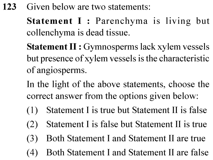
Statement I Collenchyma is a dead mechanical tissue lacking living protoplasm.
Statement II Gymnosperms lack xylem vessels in their xylem tissue.
⚡

The 30-Second Exam Hall Shortcut: ⚡ Living vs Dead Plant Tissues

⏱️ Save ~20 seconds

Collenchyma consists of living cells with localized pectin thickenings (Statement I is FALSE; Sclerenchyma is dead). Gymnosperms possess tracheids and lack true xylem vessels (Statement II is TRUE)!

1 Statement I: "Collenchyma is dead" -> False! Collenchyma is living mechanical tissue with active protoplasm (sclerenchyma is dead).
2 Statement II: "Gymnosperms lack vessels" -> True! Gymnosperm xylem consists predominantly of tracheids; vessels are absent (except in Gnetales).
3 Conclusion: Statement I is false but Statement II is true -> Option (2) / B.
🛡️
Why You Can 100% Trust This Shortcut: NCERT Biology · Anatomy of Flowering Plants (Collenchyma and Complex Tissues)
📐

Step-by-Step Rigorous Workflow & Mathematical Proof

Step 1

Evaluate Statement I (Collenchyma Properties)

Collenchyma is a simple permanent tissue consisting of living cells characterized by unevenly thickened corners due to deposition of cellulose, hemicellulose, and pectin. They possess living protoplasts and chloroplasts, providing mechanical flexibility to growing stems and petioles. Calling collenchyma "dead" is factually false (sclerenchyma is the dead mechanical tissue).

Step 2

Evaluate Statement II (Gymnosperm Xylem Properties)

In gymnosperms, xylem lacks vessels (except in order Gnetales). Water conduction occurs primarily through tracheids. Thus, the statement that gymnosperms lack vessels in their xylem is universally true in standard plant taxonomy.

Step 3

Synthesize Conclusion

Statement I is false and Statement II is true, leading directly to Option (2).

⭐

360° Option Elimination Engine Key Feature

Diagnose every choice: Know exactly why the right option works and why distractors fail.

4-Option Deep Scan
1 (1)
✗ Incorrect
Verdict & Scientific Reason:

Collenchyma is living, not dead; and gymnosperms lack vessels.

⚠️
Student Fallacy (Why candidates pick this trap): Collenchyma is living, not dead; and gymnosperms lack vessels.
2 (2)
✓ 100% Correct
Verdict & Scientific Reason:

Accurately assesses that collenchyma is a living tissue while gymnosperms lack vessels.

💡
Why this is airtight: None.
3 (3)
✗ Incorrect
Verdict & Scientific Reason:

Statement I is false because collenchyma consists of living cells.

⚠️
Student Fallacy (Why candidates pick this trap): Statement I is false because collenchyma consists of living cells.
4 (4)
✗ Incorrect
Verdict & Scientific Reason:

Statement II is factually true.

⚠️
Student Fallacy (Why candidates pick this trap): Statement II is factually true.
⚠️

NTA Trap Alert: Collenchyma vs Sclerenchyma

Confusing collenchyma with sclerenchyma because both provide mechanical support.

🛡️ How to avoid the -1 mark: Remember: Collenchyma is LIVING mechanical tissue with pectin corners; Sclerenchyma is DEAD mechanical tissue with lignified walls.
📖

Core NEET Syllabus Standard

Biology Core Botany · Anatomy of Flowering Plants Standard Benchmark
Core Scientific Principle Tested:

"Collenchyma consists of cells which are much thickened at the corners due to a deposition of cellulose, hemicellulose and pectin. Collenchymatous cells may be oval, spherical or polygonal and often contain chloroplasts... Gymnosperms lack vessels in their xylem."

Syllabus Focus: Collenchyma Living Nature and Gymnosperm Xylem Composition
✓ Core NEET UG Syllabus Anchor Syllabus & Academic Standards ↗
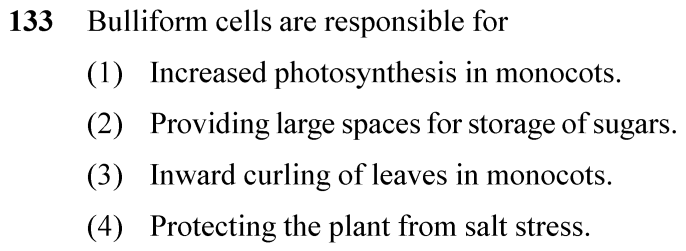
NEET 2024 · Q133 Easy
Official NEET 2024 Question 133 - Anatomy of Flowering Plants
1 Increased photosynthesis in monocots.
2 Providing large spaces for storage of sugars.
3 Inward curling of leaves in monocots. ✓ Key
4 Protecting the plant from salt stress.
💡 Verified Solution & 30-Second Hack
✓
Verified Against NTA Final Answer Key
Option (3) : Inward curling of leaves in monocots
📚 Anatomy of Flowering Plants Easy
📋 Given Parameters & Problem Setup
Cell Type Bulliform cells (motor cells)
Plant Group Monocot grasses
⚡

The 30-Second Exam Hall Shortcut: ⚡ Bulliform Cells Turgor Mechanism

⏱️ Save ~20 seconds

When water stressed, large empty bulliform cells lose turgor and become flaccid, causing leaves to curl inward to minimize surface area and transpiration!

1 Recall bulliform cell function in grass leaves:
2 - Located on the adaxial (upper) epidermis of monocot leaves.
3 - Large, empty, colourless cells.
4 - When they absorb water and are turgid, the leaf surface is exposed.
5 - When flaccid due to water stress, they cause leaves to curl inward to minimize water loss.
6 Direct match: Inward curling of leaves in monocots -> Option (3) / C.
🛡️
Why You Can 100% Trust This Shortcut: NCERT Biology · Anatomy of Flowering Plants (Isobilateral Leaf and Bulliform Cells)
📐

Step-by-Step Rigorous Workflow & Mathematical Proof

Step 1

Locate and Describe Bulliform Cells

In grasses, certain adaxial epidermal cells along the veins modify themselves into large, empty, colourless cells called bulliform cells (or motor cells).

Step 2

Understand the Turgor-Mediated Dynamic

When the bulliform cells have absorbed water and are turgid, the leaf surface is exposed. When they are flaccid due to water stress, they cause the leaves to curl inward to minimize water loss via transpiration.

Step 3

Match with Options

The primary physiological function is mediating inward curling of leaves in monocots.

⭐

360° Option Elimination Engine Key Feature

Diagnose every choice: Know exactly why the right option works and why distractors fail.

4-Option Deep Scan
1 (1)
✗ Incorrect
Verdict & Scientific Reason:

Bulliform cells are hydro-passive motor cells regulating water conservation, not halophytic salt-extruding glands.

⚠️
Student Fallacy (Why candidates pick this trap): Bulliform cells are hydro-passive motor cells regulating water conservation, not halophytic salt-extruding glands.
2 (2)
✗ Incorrect
Verdict & Scientific Reason:

Stomatal opening is controlled by the guard cells, not bulliform cells.

⚠️
Student Fallacy (Why candidates pick this trap): Stomatal opening is controlled by the guard cells, not bulliform cells.
3 (3)
✓ 100% Correct
Verdict & Scientific Reason:

Loss of turgor in bulliform cells causes leaves to curl inward to reduce exposed surface area during drought.

💡
Why this is airtight: None.
4 (4)
✗ Incorrect
Verdict & Scientific Reason:

Bulliform cells are empty, water-filled cells that do not store carbohydrates.

⚠️
Student Fallacy (Why candidates pick this trap): Bulliform cells are empty, water-filled cells that do not store carbohydrates.
⚠️

NTA Trap Alert: Stomatal Pore vs Leaf Rolling

Attributing stomatal opening/closing to bulliform cells.

🛡️ How to avoid the -1 mark: Guard cells control stomatal pores; bulliform cells control whole-leaf rolling/curling.
📖

Core NEET Syllabus Standard

Biology Core Botany · Anatomy of Flowering Plants Standard Benchmark
Core Scientific Principle Tested:

"In grasses, certain adaxial epidermal cells along the veins modify themselves into large, empty, colourless cells. These are called bulliform cells. When the bulliform cells in the leaves have absorbed water and are turgid, the leaf surface is exposed. When they are flaccid due to water stress, they make the leaves curl inwards to minimise water loss."

Syllabus Focus: Bulliform Cells and Inward Leaf Rolling in Monocots
✓ Core NEET UG Syllabus Anchor Syllabus & Academic Standards ↗
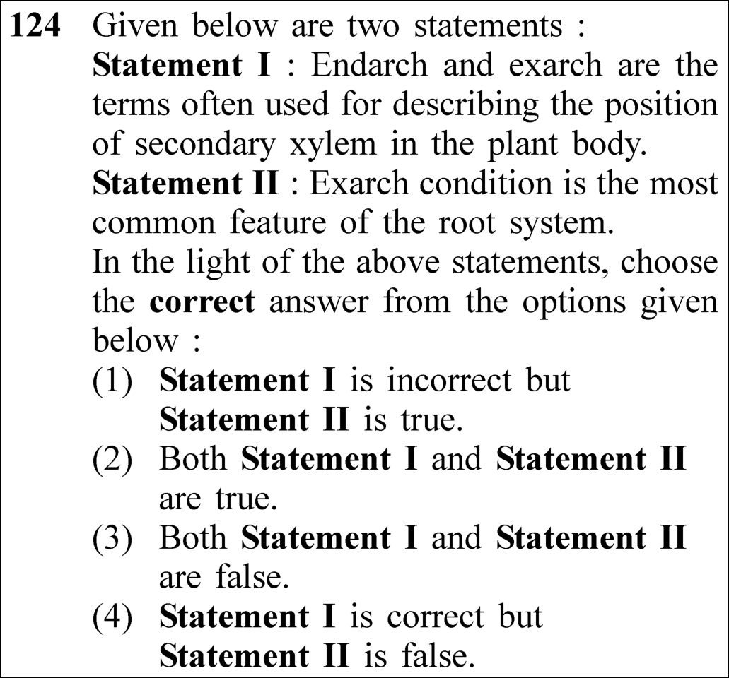
NEET 2023 · Q124 Medium
Official NEET 2023 Question 124 - Anatomy of Flowering Plants
1 Statement I is incorrect but Statement II is true. ✓ Key
2 Both Statement I and Statement II are true.
3 Both Statement I and Statement II are false.
4 Statement I is correct but Statement II is false.
💡 Verified Solution & 30-Second Hack
✓
Verified Against NTA Final Answer Key
Option (1) : Statement I is incorrect but Statement II is true.
📚 Anatomy of Flowering Plants Medium
📋 Given Parameters & Problem Setup
Statement I Endarch and exarch are terms describing position of secondary xylem.
Statement II Exarch condition is the most common feature of the root system.
⚡

The 30-Second Exam Hall Shortcut: ⚡ Endarch/Exarch = PRIMARY Xylem Only!

⏱️ Save ~25 seconds

Endarch and exarch describe the spatial orientation of protoxylem relative to metaxylem in PRIMARY xylem, NEVER secondary xylem! In roots, protoxylem is peripheral (Exarch). Statement I is false, Statement II is true!

1 Check Statement I: Endarch and exarch describe primary xylem (protoxylem & metaxylem), NOT secondary xylem -> Statement I is false/incorrect.
2 Check Statement II: In roots, protoxylem lies towards periphery and metaxylem lies towards centre (exarch) -> Statement II is true.
3 Statement I is incorrect but Statement II is true.
4 Match Option (1).
🛡️
Why You Can 100% Trust This Shortcut: Canonical NCERT Plant Anatomy xylem differentiation nomenclature.
📐

Step-by-Step Rigorous Workflow & Mathematical Proof

Step 1

Evaluation of Statement I (Primary vs. Secondary Xylem)

Endarch and exarch refer exclusively to developmental arrangements of primary xylem elements (protoxylem and metaxylem). Secondary xylem formed by vascular cambium does not exhibit endarch/exarch differentiation:

Endarch / Exarch  ⟺  Position of Protoxylem vs. Metaxylem in PRIMARY Xylem  ⟹  Statement I is Incorrect\text{\textbf{Endarch / Exarch}} \iff \text{Position of Protoxylem vs. Metaxylem in } \mathbf{\text{PRIMARY Xylem}} \implies \text{Statement I is } \mathbf{\text{Incorrect}}
Step 2

Evaluation of Statement II (Exarch Arrangement in Roots)

In stems, protoxylem lies towards pith (centre) and metaxylem towards periphery (endarch). In roots, protoxylem lies towards the periphery and metaxylem towards the centre (exarch):

Stem (Endarch):Protoxylem inner (centrifugal development)Root (Exarch):Protoxylem outer (centripetal development)  ⟹  Statement II is True\begin{aligned} \text{Stem (Endarch):} & \quad \text{Protoxylem inner (centrifugal development)} \\[3pt] \text{\textbf{Root (Exarch):}} & \quad \text{\textbf{Protoxylem outer (centripetal development)}} \implies \text{Statement II is } \mathbf{\text{True}} \end{aligned}
Step 3

Verbatim NCERT Reference

NCERT states verbatim: "In stems, the protoxylem lies towards the centre (pith) and the metaxylem lies towards the periphery of the organ. This type of primary xylem is called endarch. In roots, the protoxylem lies towards periphery and metaxylem lies towards the centre. Such arrangement of primary xylem is called exarch."

⭐

360° Option Elimination Engine Key Feature

Diagnose every choice: Know exactly why the right option works and why distractors fail.

4-Option Deep Scan
1 1
✓ 100% Correct
Verdict & Scientific Reason:

Statement I falsely refers to secondary xylem instead of primary xylem; Statement II accurately identifies exarch as typical of roots.

💡
Why this is airtight: None; rigorous anatomical precision.
2 2
✗ Incorrect
Verdict & Scientific Reason:

Statement I is false because endarch/exarch terms describe primary xylem, not secondary xylem.

⚠️
Student Fallacy (Why candidates pick this trap): Overlooking the word "secondary" in Statement I.
3 3
✗ Incorrect
Verdict & Scientific Reason:

Statement II is factually true according to plant anatomy.

⚠️
Student Fallacy (Why candidates pick this trap): Incorrectly believing roots are endarch.
4 4
✗ Incorrect
Verdict & Scientific Reason:

Both parts of this option are inverted.

⚠️
Student Fallacy (Why candidates pick this trap): Confusing stem and root xylem developmental patterns.
⚠️

NTA Trap Alert: Primary vs. Secondary Xylem Wording Trap

NTA intentionally substituted "secondary xylem" in place of "primary xylem" in Statement I. Students reading too fast recognize "endarch and exarch" and subconsciously assume it is true.

🛡️ How to avoid the -1 mark: Always verify developmental status: Endarch/Exarch applies strictly to PRIMARY xylem.
📖

Core NEET Syllabus Standard

NCERT Biology · Anatomy of Flowering Plants Standard Benchmark
Core Scientific Principle Tested:

"In roots, the protoxylem lies towards periphery and metaxylem lies towards the centre. Such arrangement of primary xylem is called exarch."

Syllabus Focus: Complex Tissues (Xylem)
✓ Core NEET UG Syllabus Anchor Syllabus & Academic Standards ↗
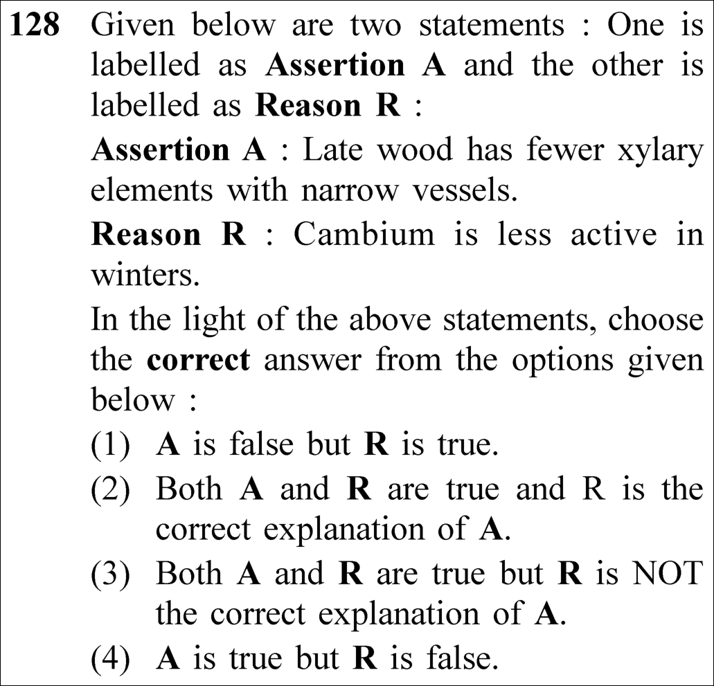
NEET 2023 · Q128 Medium
Official NEET 2023 Question 128 - Anatomy of Flowering Plants
1 A is false but R is true.
2 Both A and R are true and R is the correct explanation of A. ✓ Key
3 Both A and R are true but R is NOT the correct explanation of A.
4 A is true but R is false.
💡 Verified Solution & 30-Second Hack
✓
Verified Against NTA Final Answer Key
Option (2) : Both A and R are true and R is the correct explanation of A.
📚 Anatomy of Flowering Plants Medium
📋 Given Parameters & Problem Setup
Assertion (A) Late wood has fewer xylary elements with narrow vessels.
Reason (R) Cambium is less active in winters.
⚡

The 30-Second Exam Hall Shortcut: ⚡ Seasonal Cambial Activity: Winter Sluggishness = Late Wood

⏱️ Save ~25 seconds

In winter/autumn, vascular cambium is less active, forming fewer xylem elements with narrow lumens (late/autumn wood). In spring, cambium is highly active, producing numerous elements with wide vessels (spring/early wood)!

1 Assertion: Late wood (autumn wood) has fewer xylary elements with narrow vessels -> True.
2 Reason: In winter, climatic conditions are adverse and cambium is less active -> True.
3 Causal link: The reduced cambial activity in winter directly causes the formation of fewer elements with narrow vessels.
4 R is the correct explanation of A -> Select Option (2) Both A and R are true and R is the correct explanation of A.
🛡️
Why You Can 100% Trust This Shortcut: Canonical NCERT Plant Anatomy seasonal cambial kinetics.
📐

Step-by-Step Rigorous Workflow & Mathematical Proof

Step 1

Physiological Basis of Annual Rings

Vascular cambium activity is modulated by seasonal climatic fluctuations in temperate regions:

Spring Wood (Early Wood):Cambium highly active  ⟹  Many xylary elements with wide vessels (low density)Late Wood (Autumn Wood):Cambium less active in winter  ⟹  Fewer xylary elements with narrow vessels (high density)\begin{aligned} \text{\textbf{Spring Wood (Early Wood):}} & \quad \text{Cambium highly active} \implies \text{Many xylary elements with } \mathbf{\text{wide vessels (low density)}} \\[4pt] \text{\textbf{Late Wood (Autumn Wood):}} & \quad \mathbf{\text{Cambium less active in winter}} \implies \mathbf{\text{Fewer xylary elements with narrow vessels (high density)}} \end{aligned}
Step 2

Verbatim NCERT Reference

NCERT states verbatim: "In winter, the cambium is less active and forms fewer xylary elements that have narrow vessels, and this wood is called autumn wood or late wood."

Step 3

Assertion-Reason Causal Validation

The diminished mitotic rate of cambial fusiform initials during winter is the direct physiological cause for the anatomical traits (fewer elements, narrow vessels) of late wood. Hence, Reason R correctly explains Assertion A.

⭐

360° Option Elimination Engine Key Feature

Diagnose every choice: Know exactly why the right option works and why distractors fail.

4-Option Deep Scan
1 1
✗ Incorrect
Verdict & Scientific Reason:

Assertion is factually true: late wood is characterized by narrow vessels and fewer elements.

⚠️
Student Fallacy (Why candidates pick this trap): Confusing early wood wide vessels with late wood narrow vessels.
2 2
✓ 100% Correct
Verdict & Scientific Reason:

Both statements are true and Reason provides the exact physiological cause for Assertion.

💡
Why this is airtight: None; rigorous physiological deduction.
3 3
✗ Incorrect
Verdict & Scientific Reason:

Cambial activity directly controls xylary element number and vessel diameter; the link is directly causal.

⚠️
Student Fallacy (Why candidates pick this trap): Failing to recognize cambial activity as the direct driver of xylem morphology.
4 4
✗ Incorrect
Verdict & Scientific Reason:

Reason is factually true: cambium is less active in winters in temperate zones.

⚠️
Student Fallacy (Why candidates pick this trap): Assuming cambial activity remains constant year-round.
⚠️

NTA Trap Alert: Spring vs. Autumn Wood Property Reversal Trap

Students frequently mix up the traits: Early/Spring wood is light in color with low density and wide vessels; Late/Autumn wood is dark in color with high density and narrow vessels.

🛡️ How to avoid the -1 mark: Spring = Vigorous growth (wide vessels, high activity). Winter/Late = Dormancy (narrow vessels, low activity).
📖

Core NEET Syllabus Standard

NCERT Biology · Anatomy of Flowering Plants Standard Benchmark
Core Scientific Principle Tested:

"In winter, the cambium is less active and forms fewer xylary elements that have narrow vessels, and this wood is called autumn wood or late wood."

Syllabus Focus: Secondary Growth (Spring Wood and Autumn Wood)
✓ Core NEET UG Syllabus Anchor Syllabus & Academic Standards ↗
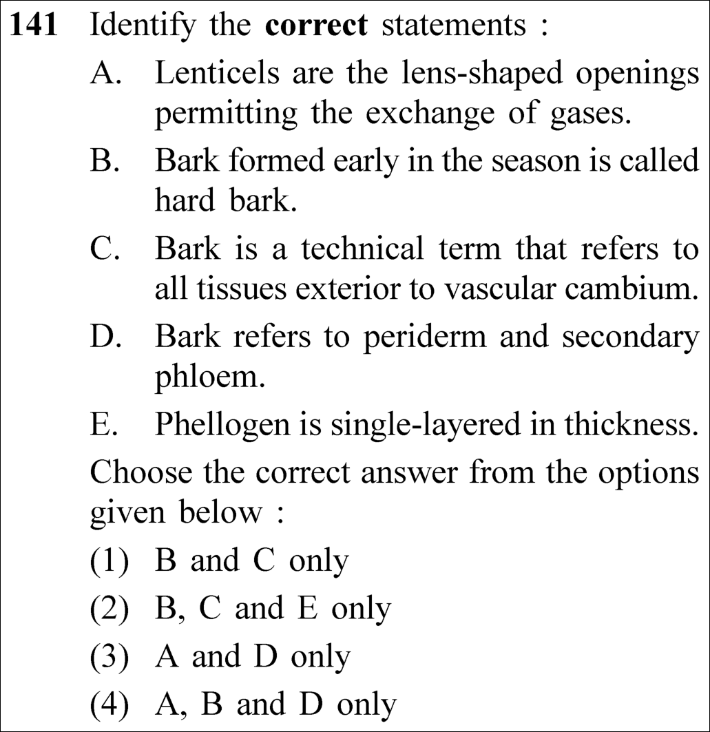
NEET 2023 · Q141 Medium
Official NEET 2023 Question 141 - Anatomy of Flowering Plants
1 B and C only
2 B, C and E only
3 A and D only ✓ Key
4 A, B and D only
💡 Verified Solution & 30-Second Hack
✓
Verified Against NTA Final Answer Key
Option (3) : A and D only
📚 Anatomy of Flowering Plants Medium
📋 Given Parameters & Problem Setup
Anatomical Statements 5 statements (A-E) on periderm, lenticels, phellogen, and bark
⚡

The 30-Second Exam Hall Shortcut: ⚡ Bark Technicality & Seasonal Texture Precision

⏱️ Save ~30 seconds

Spot the false statements: Early bark is SOFT (not hard, so B is false). Bark is a NON-TECHNICAL term (not technical, so C is false). Phellogen is a COUPLE OF LAYERS thick (not single-layered, so E is false). Thus, ONLY A and D are correct!

1 Statement A: Lenticels are lens-shaped openings permitting gas exchange -> True.
2 Statement B: Bark formed early is hard bark -> False (Early bark is soft bark; late bark is hard bark).
3 Statement C: Bark is a technical term -> False (NCERT: "Bark is a non-technical term referring to all tissues exterior to vascular cambium").
4 Statement D: Bark refers to periderm and secondary phloem -> True.
5 Statement E: Phellogen is single-layered -> False (Phellogen is a couple of layers thick).
6 A and D only -> Select Option (3).
🛡️
Why You Can 100% Trust This Shortcut: Canonical NCERT Plant Anatomy periderm and secondary phloem technical definitions.
📐

Step-by-Step Rigorous Workflow & Mathematical Proof

Step 1

Evaluation of Statements A and D (Valid Statements)

Analyzing the veracity of anatomical statements according to NCERT:

Statement A:Lenticels are lens-shaped aerating pores in bark  ⟹  TRUEStatement D:Bark anatomically includes periderm + secondary phloem  ⟹  TRUE\begin{aligned} \text{\textbf{Statement A:}} & \quad \text{Lenticels are lens-shaped aerating pores in bark} \implies \mathbf{\text{TRUE}} \\[3pt] \text{\textbf{Statement D:}} & \quad \text{Bark anatomically includes periderm } + \text{ secondary phloem} \implies \mathbf{\text{TRUE}} \end{aligned}
Step 2

Evaluation of Statements B, C, and E (Invalid Statements)

Analyzing the specific errors planted in the distractors:

Statement B:Bark formed early in season is soft bark, NOT hard bark  ⟹  FALSEStatement C:Bark is a non-technical term, NOT a technical term  ⟹  FALSEStatement E:Phellogen is a couple of layers thick, NOT single-layered  ⟹  FALSE\begin{aligned} \text{\textbf{Statement B:}} & \quad \text{Bark formed early in season is } \mathbf{\text{soft bark}}, \text{ NOT hard bark} \implies \mathbf{\text{FALSE}} \\[3pt] \text{\textbf{Statement C:}} & \quad \text{Bark is a } \mathbf{\text{non-technical term}}, \text{ NOT a technical term} \implies \mathbf{\text{FALSE}} \\[3pt] \text{\textbf{Statement E:}} & \quad \text{Phellogen is } \mathbf{\text{a couple of layers thick}}, \text{ NOT single-layered} \implies \mathbf{\text{FALSE}} \end{aligned}
Step 3

Conclusion

Only Statements A and D are factually and technically correct. Option (3) is the required answer.

⭐

360° Option Elimination Engine Key Feature

Diagnose every choice: Know exactly why the right option works and why distractors fail.

4-Option Deep Scan
1 1
✗ Incorrect
Verdict & Scientific Reason:

Both B (early bark is soft, not hard) and C (bark is a non-technical term) are false.

⚠️
Student Fallacy (Why candidates pick this trap): Failing to catch the single-word negative distortions in B and C.
2 2
✗ Incorrect
Verdict & Scientific Reason:

All three statements B, C, and E are false.

⚠️
Student Fallacy (Why candidates pick this trap): Selecting all the erroneous statements.
3 3
✓ 100% Correct
Verdict & Scientific Reason:

Statements A and D are completely accurate according to NCERT textbook definitions.

💡
Why this is airtight: None; rigorous critical analysis.
4 4
✗ Incorrect
Verdict & Scientific Reason:

Includes false statement B.

⚠️
Student Fallacy (Why candidates pick this trap): Overlooking that early-season bark is soft bark.
⚠️

NTA Trap Alert: Technical vs. Non-Technical Bark Trap

NCERT explicitly states: "Bark is a non-technical term...". NTA frequently removes the "non-" to create a deceptive distractor that sounds authoritative.

🛡️ How to avoid the -1 mark: Remember: Bark is NON-TECHNICAL. Early bark is SOFT. Phellogen is A COUPLE OF LAYERS thick.
📖

Core NEET Syllabus Standard

NCERT Biology · Anatomy of Flowering Plants Standard Benchmark
Core Scientific Principle Tested:

"Lenticels are lens-shaped openings permitting exchange of gases. Bark is a non-technical term referring to periderm and secondary phloem."

Syllabus Focus: Secondary Growth (Cork Cambium and Bark)
✓ Core NEET UG Syllabus Anchor Syllabus & Academic Standards ↗
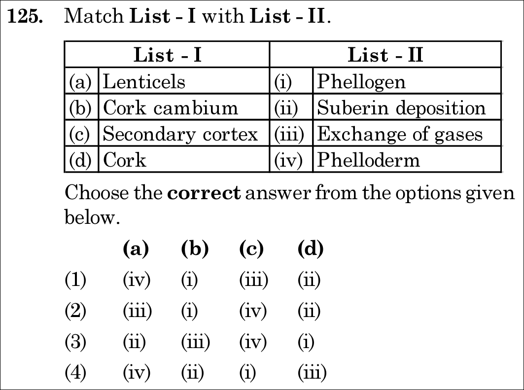
NEET 2021 · Q125 Easy
Official NEET 2021 Question 125 - Anatomy of Flowering Plants
1 (iv) (i) (iii) (ii)
2 (iii) (i) (iv) (ii) ✓ Key
3 (ii) (iii) (iv) (i)
4 (iv) (ii) (i) (iii)
💡 Verified Solution & 30-Second Hack
✓
Verified Against NTA Final Answer Key
Option (2) : (a)-(iii), (b)-(i), (c)-(iv), (d)-(ii)
📚 Anatomy of Flowering Plants Easy
📋 Given Parameters & Problem Setup
List I (a) Lenticels, (b) Cork cambium, (c) Secondary cortex, (d) Cork
List II (i) Phellogen, (ii) Suberin deposition, (iii) Exchange of gases, (iv) Phelloderm
⚡

The 30-Second Exam Hall Shortcut: ⚡ Classic Periderm Triad: Phellogen (Cork Cambium) & Phelloderm (Secondary Cortex)

⏱️ Save ~20 seconds

Cork cambium = Phellogen (b-i); Secondary cortex = Phelloderm (c-iv); Cork = Phellem with Suberin (d-ii); Lenticels = Gas exchange openings in bark (a-iii). This immediately gives (a)-(iii), (b)-(i), (c)-(iv), (d)-(ii)   ⟹  \implies Option (2)!

1 Lenticels: Lens-shaped openings in bark for aerating internal tissues   ⟹  \implies (a)-(iii).
2 Cork cambium: The secondary lateral meristem termed Phellogen   ⟹  \implies (b)-(i).
3 Secondary cortex: Parenchymatous cells cut off toward the inside, termed Phelloderm   ⟹  \implies (c)-(iv).
4 Cork: Dead cells with suberized walls cut off to the outside (Phellem)   ⟹  \implies (d)-(ii).
5 Match Option (2).
🛡️
Why You Can 100% Trust This Shortcut: Canonical NCERT Anatomy of Flowering Plants on Secondary Growth and Periderm.
📐

Step-by-Step Rigorous Workflow & Mathematical Proof

Step 1

Anatomy of the Periderm Complex

During secondary growth in dicot stems, the increase in girth tears the outer cortical and epidermal layers. A secondary lateral meristem arises in the cortex:

Cork Cambium (Phellogen):Secondary meristem developing in cortex   ⟹  (b)-(i)Cork (Phellem):Cells cut off to the outside with suberized walls   ⟹  (d)-(ii)Secondary Cortex (Phelloderm):Parenchymatous cells cut off to the inside   ⟹  (c)-(iv)Periderm:Phellogen+Phellem+Phelloderm\begin{aligned} \text{\textbf{Cork Cambium (Phellogen):}} & \quad \text{Secondary meristem developing in cortex } \implies \text{(b)-(i)} \\[4pt] \text{\textbf{Cork (Phellem):}} & \quad \text{Cells cut off to the outside with suberized walls } \implies \text{(d)-(ii)} \\[4pt] \text{\textbf{Secondary Cortex (Phelloderm):}} & \quad \text{Parenchymatous cells cut off to the inside } \implies \text{(c)-(iv)} \\[4pt] \text{\textbf{Periderm:}} & \quad \text{Phellogen} + \text{Phellem} + \text{Phelloderm} \end{aligned}
Step 2

Structure and Function of Lenticels

At certain regions, the phellogen cuts off closely arranged parenchymatous complementary cells instead of cork cells. These cells rupture the epidermis, forming lens-shaped apertures called lenticels that permit exchange of gases between the atmosphere and internal cortical tissues   ⟹  \implies (a)-(iii).

⭐

360° Option Elimination Engine Key Feature

Diagnose every choice: Know exactly why the right option works and why distractors fail.

4-Option Deep Scan
1 1
✗ Incorrect
Verdict & Scientific Reason:

Mismatches Lenticels with phelloderm and Secondary cortex with gas exchange.

⚠️
Student Fallacy (Why candidates pick this trap): Confusing the gas exchange aperture with the inner cortical layer.
2 2
✓ 100% Correct
Verdict & Scientific Reason:

All four periderm and bark structures are matched flawlessly to their anatomical identities.

💡
Why this is airtight: None. Accurate pairing.
3 3
✗ Incorrect
Verdict & Scientific Reason:

Mismatches Lenticels with suberin and Cork cambium with gas exchange.

⚠️
Student Fallacy (Why candidates pick this trap): Swapping lenticels with cork suberin.
4 4
✗ Incorrect
Verdict & Scientific Reason:

Gross misassignment of all anatomical terms.

⚠️
Student Fallacy (Why candidates pick this trap): Random guessing among matching options.
⚠️

NTA Trap Alert: Phellogen, Phellem, and Phelloderm Nomenclature Mix-Up

The Greek roots are easily inverted: Phellogen = Genesis/Origin (Cork cambium); Phellem = Cork; Phelloderm = Dermis/Cortex (Secondary cortex).

🛡️ How to avoid the -1 mark: Mnemonic: "Phellogen Generates; Phellem is Suberin film; Phelloderm is Deep cortex".
📖

Core NEET Syllabus Standard

NEET / NCERT Biology · Anatomy of Flowering Plants Standard Benchmark
Core Scientific Principle Tested:

"Phellogen, phellem, and phelloderm are collectively known as periderm. Lenticels permit the exchange of gases between the outer atmosphere and the internal tissue."

Syllabus Focus: Secondary Growth: Cork Cambium and Lenticels
✓ Core NEET UG Syllabus Anchor Syllabus & Academic Standards ↗
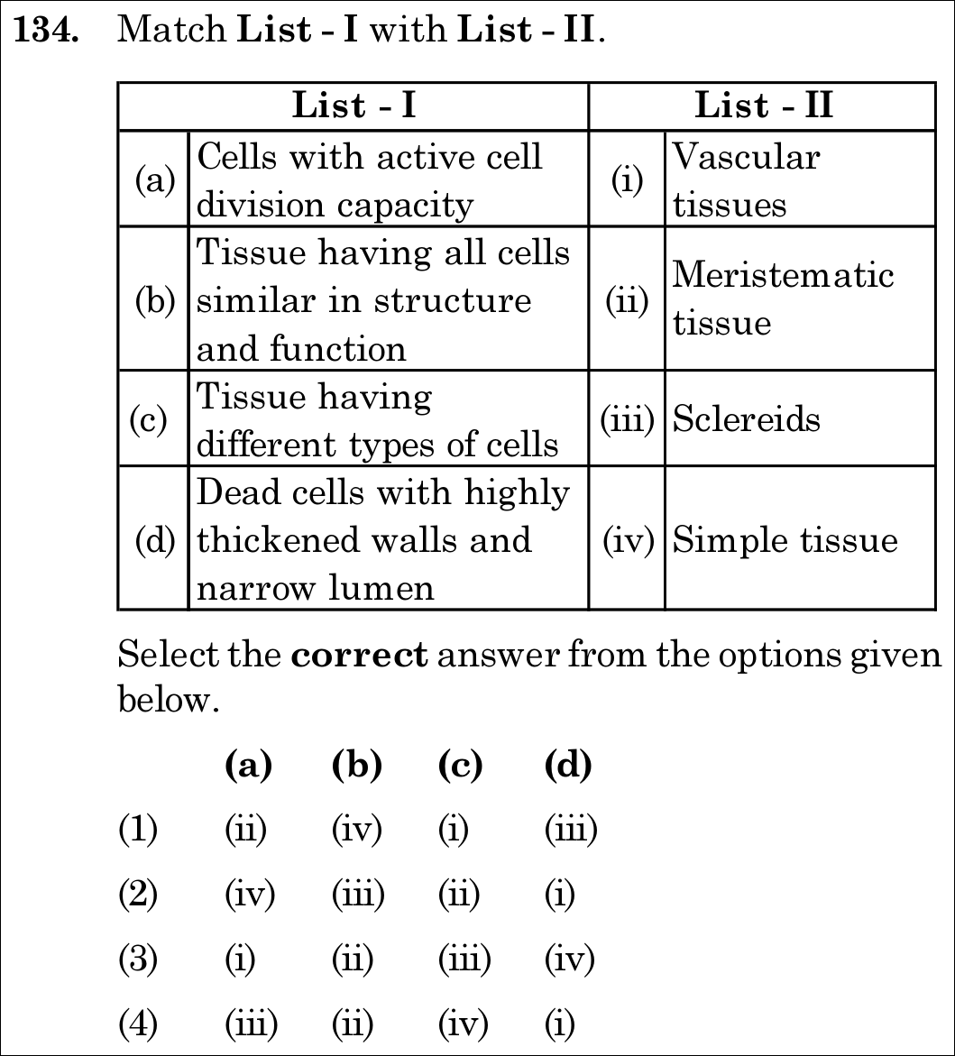
NEET 2021 · Q134 Easy
Official NEET 2021 Question 134 - Anatomy of Flowering Plants
1 (ii) (iv) (i) (iii) ✓ Key
2 (iv) (iii) (ii) (i)
3 (i) (ii) (iii) (iv)
4 (iii) (ii) (iv) (i)
💡 Verified Solution & 30-Second Hack
✓
Verified Against NTA Final Answer Key
Option (1) : (a)-(ii), (b)-(iv), (c)-(i), (d)-(iii)
📚 Anatomy of Flowering Plants Easy
📋 Given Parameters & Problem Setup
List I (a) Cells with active cell division capacity, (b) Tissue having all cells similar in structure and function, (c) Tissue having different types of cells, (d) Dead cells with highly thickened walls and narrow lumen
List II (i) Vascular tissues, (ii) Meristematic tissue, (iii) Sclereids, (iv) Simple tissue
⚡

The 30-Second Exam Hall Shortcut: ⚡ Fast Pairing: Active Division = Meristem (ii) & Dead Thick Lumen = Sclereids (iii)

⏱️ Save ~20 seconds

Active cell division = Meristematic tissue (a-ii); Homogeneous cells similar in structure = Simple tissue (b-iv); Heterogeneous complex cells = Vascular tissue (c-i); Highly thickened dead cells with narrow lumen = Sclereids (d-iii). Matches Option (1)!

1 (a) Active cell division capacity   ⟹  \implies (ii) Meristematic tissue.
2 (b) All cells similar in structure and function   ⟹  \implies (iv) Simple tissue (parenchyma, collenchyma, sclerenchyma).
3 (c) Tissue composed of different cell types   ⟹  \implies (i) Vascular tissues (complex tissues: xylem and phloem).
4 (d) Dead cells with thick lignified walls and narrow lumen   ⟹  \implies (iii) Sclereids (sclerenchyma).
5 Match Option (1).
🛡️
Why You Can 100% Trust This Shortcut: Canonical NCERT Anatomy of Flowering Plants on Meristems and Permanent Tissues.
📐

Step-by-Step Rigorous Workflow & Mathematical Proof

Step 1

Histological Classification of Plant Tissues

Plant tissues are systematically organized into meristematic and permanent tissues based on division capability and cellular uniformity:

(a) Cells with active mitotic division⟷(ii) Meristematic tissue(b) All cells identical in structure and function⟷(iv) Simple tissue (e.g., parenchyma)(c) Multiple distinct cell types working as a unit⟷(i) Vascular tissues (complex xylem/phloem)(d) Dead cells with lignified walls and narrow lumen⟷(iii) Sclereids (stone cells/grit cells)\begin{aligned} \text{(a) Cells with active mitotic division} & \longleftrightarrow \text{(ii) Meristematic tissue} \\[3pt] \text{(b) All cells identical in structure and function} & \longleftrightarrow \text{(iv) Simple tissue (e.g., parenchyma)} \\[3pt] \text{(c) Multiple distinct cell types working as a unit} & \longleftrightarrow \text{(i) Vascular tissues (complex xylem/phloem)} \\[3pt] \text{(d) Dead cells with lignified walls and narrow lumen} & \longleftrightarrow \text{(iii) Sclereids (stone cells/grit cells)} \end{aligned}
Step 2

Option Alignment and Verification

Checking (a)-(ii) narrows down candidates to Option (1). Verifying (b)-(iv), (c)-(i), and (d)-(iii) confirms complete 100% agreement with Option (1).

⭐

360° Option Elimination Engine Key Feature

Diagnose every choice: Know exactly why the right option works and why distractors fail.

4-Option Deep Scan
1 1
✓ 100% Correct
Verdict & Scientific Reason:

Flawless correspondence according to canonical plant histology definitions.

💡
Why this is airtight: None. Clean first-principles match.
2 2
✗ Incorrect
Verdict & Scientific Reason:

Mismatches active division with simple tissue and dead cells with vascular tissue.

⚠️
Student Fallacy (Why candidates pick this trap): Confusing meristematic division with simple tissue.
3 3
✗ Incorrect
Verdict & Scientific Reason:

Mismatches dividing cells with vascular tissues.

⚠️
Student Fallacy (Why candidates pick this trap): Conflating active growth with vascular sap conduction.
4 4
✗ Incorrect
Verdict & Scientific Reason:

Inverts dividing cells with sclereids.

⚠️
Student Fallacy (Why candidates pick this trap): Swapping meristematic tissue with dead sclereids.
⚠️

NTA Trap Alert: Simple Tissue vs Complex Vascular Tissue Definitions

NCERT defines Simple Tissue as "made of only one type of cells" and Complex Tissue as "made of more than one type of cells" (vascular tissues).

🛡️ How to avoid the -1 mark: One type = Simple; Multiple types = Complex / Vascular.
📖

Core NEET Syllabus Standard

NEET / NCERT Biology · Anatomy of Flowering Plants Standard Benchmark
Core Scientific Principle Tested:

"Meristems possess active dividing capacity; simple tissues have similar cells; complex/vascular tissues have heterogeneous cells; sclereids are thick-walled dead cells with narrow lumen."

Syllabus Focus: The Tissues: Meristematic, Simple, and Complex Permanent Tissues
✓ Core NEET UG Syllabus Anchor Syllabus & Academic Standards ↗
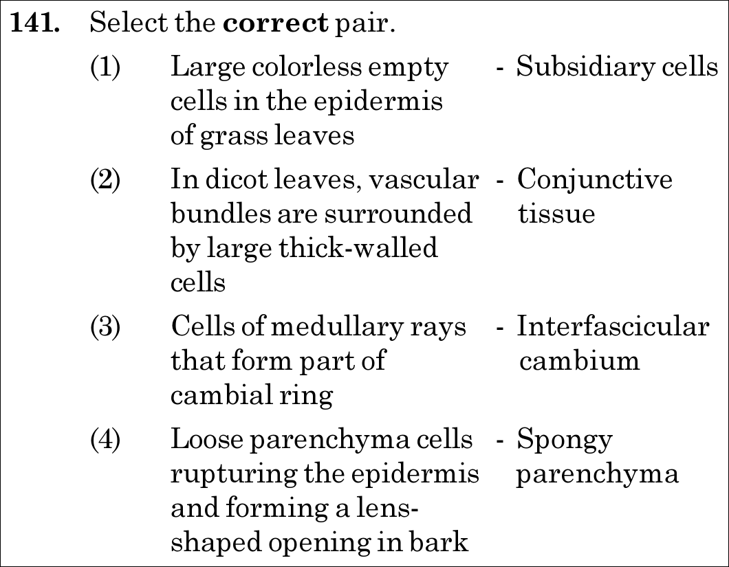
NEET 2021 · Q141 Easy
Official NEET 2021 Question 141 - Anatomy of Flowering Plants
1 Large colorless empty - Subsidiary cells cells in the epidermis of grass leaves
2 In dicot leaves, vascular - Conjunctive bundles are surrounded tissue by large thick-walled cells
3 Cells of medullary rays - Interfascicular that form part of cambium cambial ring ✓ Key
4 Loose parenchyma cells - Spongy rupturing the epidermis parenchyma and forming a lens- shaped opening in bark
💡 Verified Solution & 30-Second Hack
✓
Verified Against NTA Final Answer Key
Option (3) : Cells of medullary rays that form part of cambial ring - Interfascicular cambium
📚 Anatomy of Flowering Plants Easy
📋 Given Parameters & Problem Setup
Anatomical Pairs Four structure-function / origin pairings in plant anatomy
Target Select the CORRECT pair
⚡

The 30-Second Exam Hall Shortcut: ⚡ Medullary Rays →\to Interfascicular Cambium (Classic Dedifferentiation!)

⏱️ Save ~20 seconds

In dicot stems, the cells of medullary rays adjoining intrafascicular cambium become meristematic (dedifferentiate) and form the interfascicular cambium, completing the continuous cambial ring! Pair (3) is 100% correct. Match Option (3)!

1 Check Pair (1): Large colorless empty epidermal cells in grasses are Bulliform cells, NOT subsidiary cells   ⟹  \implies INCORRECT.
2 Check Pair (2): In dicot leaves, vascular bundles are surrounded by bundle sheath cells, NOT conjunctive tissue   ⟹  \implies INCORRECT.
3 Check Pair (3): Cells of medullary rays forming part of cambial ring = Interfascicular cambium   ⟹  \implies CORRECT verbatim NCERT.
4 Check Pair (4): Loose parenchyma cells rupturing epidermis to form lens-shaped opening = Complementary cells / Lenticels, NOT spongy parenchyma   ⟹  \implies INCORRECT.
5 Match Option (3).
🛡️
Why You Can 100% Trust This Shortcut: Canonical NCERT Anatomy of Flowering Plants on Cambial Ring Formation.
📐

Step-by-Step Rigorous Workflow & Mathematical Proof

Step 1

Formation of the Complete Cambial Ring in Dicot Stems

In young dicot stems, cambium present between primary xylem and primary phloem within a vascular bundle is the intrafascicular cambium (primary lateral meristem):

Intrafascicular Cambium+Dedifferentiated Medullary Ray Cells⏟Interfascicular Cambium⟶Continuous Vascular Cambial Ring\text{Intrafascicular Cambium} + \underbrace{\text{Dedifferentiated Medullary Ray Cells}}_{\mathbf{\text{Interfascicular Cambium}}} \longrightarrow \mathbf{\text{Continuous Vascular Cambial Ring}}
Step 2

Deconstruction and Refutation of Incorrect Pairs

- Pair (1): Large, empty, colorless epidermal cells on the adaxial surface of grass leaves are bulliform (motor) cells, which control leaf rolling to minimize transpiration (subsidiary cells flank stomatal guard cells). - Pair (2): In leaves, vascular bundles are encased by a layer of tightly packed cells known as the bundle sheath (conjunctive tissue is the parenchymatous tissue between xylem and phloem in roots). - Pair (4): The loose, thin-walled parenchymatous cells produced by phellogen that rupture the epidermis are complementary cells, forming lenticels (spongy parenchyma is the loosely arranged mesophyll tissue in leaves).

⭐

360° Option Elimination Engine Key Feature

Diagnose every choice: Know exactly why the right option works and why distractors fail.

4-Option Deep Scan
1 1
✗ Incorrect
Verdict & Scientific Reason:

Incorrect. These specialized cells are bulliform cells (motor cells), not subsidiary cells.

⚠️
Student Fallacy (Why candidates pick this trap): Confusing stomatal subsidiary cells with bulliform cells.
2 2
✗ Incorrect
Verdict & Scientific Reason:

Incorrect. Vascular bundles in leaves are surrounded by bundle sheath cells; conjunctive tissue is found in roots.

⚠️
Student Fallacy (Why candidates pick this trap): Confusing leaf bundle sheath with root conjunctive tissue.
3 3
✓ 100% Correct
Verdict & Scientific Reason:

This pair is completely TRUE. Cells of medullary rays adjoining fascicular cambium dedifferentiate to form interfascicular cambium.

💡
Why this is airtight: None. Canonical textbook relationship.
4 4
✗ Incorrect
Verdict & Scientific Reason:

Incorrect. These are complementary cells of lenticels, not leaf spongy parenchyma.

⚠️
Student Fallacy (Why candidates pick this trap): Conflating aerating bark complementary cells with leaf spongy mesophyll.
⚠️

NTA Trap Alert: Intrafascicular vs Interfascicular Cambium Origin

Intrafascicular cambium is primary in origin (present in primary bundle); Interfascicular cambium is secondary in origin, formed from medullary ray cells by dedifferentiation.

🛡️ How to avoid the -1 mark: Inter = Between bundles   ⟹  \implies formed by medullary rays between the vascular bundles.
📖

Core NEET Syllabus Standard

NEET / NCERT Biology · Anatomy of Flowering Plants Standard Benchmark
Core Scientific Principle Tested:

"In dicot stems, the cells of cambium present between primary xylem and primary phloem is the intrafascicular cambium. The cells of medullary rays, adjoining these intrafascicular cambium become active and form the interfascicular cambium."

Syllabus Focus: Secondary Growth: Vascular Cambium
✓ Core NEET UG Syllabus Anchor Syllabus & Academic Standards ↗
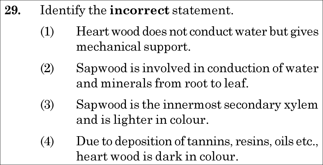
NEET 2020 · Q29 Easy
Official NEET 2020 Question 29 - Anatomy of Flowering Plants
1 Heart wood does not conduct water but gives mechanical support.
2 Sapwood is involved in conduction of water and minerals from root to leaf.
3 Sapwood is the innermost secondary xylem and is lighter in colour. ✓ Key
4 Due to deposition of tannins, resins, oils etc., heart wood is dark in colour.
💡 Verified Solution & 30-Second Hack
✓
Verified Against NTA Final Answer Key
Option (3) : Sapwood is the innermost secondary xylem and is lighter in colour.
📚 Anatomy of Flowering Plants Easy
📋 Given Parameters & Problem Setup
Tissue Types Heartwood (Duramen) and Sapwood (Alburnum)
Question Objective Identify the INCORRECT statement
⚡

The 30-Second Exam Hall Shortcut: ⚡ "HEARTwood is the INNMOST Dark Wood; SAPwood is the PERIPHERAL Light Wood!"

⏱️ Save ~20 seconds

Heartwood occupies the central innermost core of secondary xylem (dark, dead, plugged with tyloses for mechanical support). Sapwood is the PERIPHERAL (outer) secondary xylem (light-colored, active in water conduction). Claiming sapwood is "innermost" is completely FALSE! Select Option (3).

1 Recall spatial zones of secondary xylem in an old tree trunk: - Innermost central region = Heartwood (Duramen): Non-conducting, dark brown, plugged with gums/resins/tannins, gives mechanical support. - Peripheral outer region = Sapwood (Alburnum): Water-conducting, lighter in colour.
2 Analyze Statement (3): Claims "Sapwood is the innermost secondary xylem" -> Blatantly false!
3 Statement (3) is incorrect and therefore the correct answer.
🛡️
Why You Can 100% Trust This Shortcut: NCERT Biology · Anatomy of Flowering Plants (Secondary Growth - Heartwood and Sapwood: In old trees, the greater part of secondary xylem is dark brown due to deposition of organic compounds... This region comprises dead elements with highly lignified walls and is called heartwood. The peripheral region of the secondary xylem, is lighter in colour and is known as the sapwood. It is involved in the conduction of water and minerals from root to leaf).
📐

Step-by-Step Rigorous Workflow & Mathematical Proof

Step 1

Anatomy and Topography of Secondary Xylem in Old Woody Stems

In arborescent dicotyledonous trees undergoing multi-year secondary growth, the secondary xylem differentiates into two concentric zones: - Heartwood (Duramen): The innermost, central cylinder of secondary xylem. Tracheids and vessels become non-functional and plugged by balloon-like parenchymatous ingrowths called tyloses. Deposition of aromatic tannins, resins, oils, gums, and essential oils imparts a dark brown or black colour, durable decay-resistance, and massive mechanical tensile strength. It does not conduct water. - Sapwood (Alburnum): The peripheral, outer region of secondary xylem. It contains living xylem parenchyma and unobstructed tracheary elements that are actively involved in the conduction of sap (water and dissolved mineral salts) from roots to leaves. It is distinctly lighter in colour.

{Heartwood (Duramen):Innermost central region, Dark, Dead, Mechanical SupportSapwood (Alburnum):Peripheral outer region, Lighter colour, Water Conduction\begin{cases} \textbf{Heartwood (Duramen):} & \mathbf{Innermost\,central\,region}, \, \text{Dark, Dead, Mechanical Support} \\[4pt] \textbf{Sapwood (Alburnum):} & \mathbf{Peripheral\,outer\,region}, \, \text{Lighter colour, Water Conduction} \end{cases}
Step 2

Resolution of the False Statement

Statement (3) claims that sapwood is the "innermost secondary xylem". Because sapwood is strictly peripheral, statement (3) is anatomically false and is the required answer.

⭐

360° Option Elimination Engine Key Feature

Diagnose every choice: Know exactly why the right option works and why distractors fail.

4-Option Deep Scan
1 A
✗ Incorrect
Verdict & Scientific Reason:

This is a true statement. Heartwood vessels are occluded by tyloses and provide structural support.

⚠️
Student Fallacy (Why candidates pick this trap): Assuming heartwood conducts water.
2 B
✗ Incorrect
Verdict & Scientific Reason:

This is a true statement describing the primary physiological role of sapwood.

⚠️
Student Fallacy (Why candidates pick this trap): Confusing sapwood with heartwood.
3 C
✓ 100% Correct
Verdict & Scientific Reason:

Correct as the incorrect statement. Sapwood is the peripheral secondary xylem, not the innermost.

💡
Why this is airtight: None. Direct recall of wood zonal topography.
4 D
✗ Incorrect
Verdict & Scientific Reason:

This is a true statement explaining the dark pigmentation and decay resistance of heartwood.

⚠️
Student Fallacy (Why candidates pick this trap): Failing to realize that organic deposits darken heartwood.
⚠️

NTA Trap Alert: Innermost vs Peripheral Position Inversion Trap

Students frequently remember that sapwood is light and heartwood is dark, but rush past the spatial term: Heartwood is at the "heart" (center/innermost) of the tree; Sapwood is peripheral (outer).

🛡️ How to avoid the -1 mark: Use the anatomical root: The "Heart" of a tree is in the CENTER/INNERMOST -> Heartwood is innermost.
📖

Core NEET Syllabus Standard

NEET / NCERT Biology · Anatomy of Flowering Plants Standard Benchmark
Core Scientific Principle Tested:

"Heartwood is the central/innermost secondary xylem; sapwood is the peripheral conducting secondary xylem."

Syllabus Focus: Heartwood and Sapwood
✓ Core NEET UG Syllabus Anchor Syllabus & Academic Standards ↗
NEET 2020 · Q36 Easy
Official NEET 2020 Question 36 - Anatomy of Flowering Plants
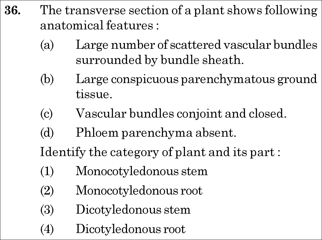
1 Monocotyledonous stem ✓ Key
2 Monocotyledonous root
3 Dicotyledonous stem
4 Dicotyledonous root
💡 Verified Solution & 30-Second Hack
✓
Verified Against NTA Final Answer Key
Option (1) : Monocotyledonous stem
📚 Anatomy of Flowering Plants Easy
📋 Given Parameters & Problem Setup
Feature (a) Large number of scattered vascular bundles surrounded by bundle sheath
Feature (b) Large conspicuous parenchymatous ground tissue
Feature (c) Vascular bundles conjoint and closed
Feature (d) Phloem parenchyma absent
⚡

The 30-Second Exam Hall Shortcut: ⚡ "Scattered + Closed + Phloem Parenchyma Absent = Monocot Stem!"

⏱️ 35 seconds

Roots always have radial bundles. Dicot stems have ringed, open bundles with cambium. Stems with scattered, closed bundles (atactostele) lacking phloem parenchyma uniquely describe a Monocotyledonous Stem! Mark Option (1) instantly.

1 Stems have conjoint vascular bundles; roots have radial bundles (eliminates monocot/dicot roots).
2 Scattered vascular bundles (atactostele) + closed bundles (no cambium) + absence of phloem parenchyma = Monocot stem.
3 Select Option (1) Monocotyledonous stem.
🛡️
Why You Can 100% Trust This Shortcut: NCERT Biology · Anatomy of Flowering Plants (Monocotyledonous Stem)
📐

Step-by-Step Rigorous Workflow & Mathematical Proof

Step 1

Eliminate Root Anatomy

In all roots (dicot and monocot), vascular bundles are arranged radially (xylem and phloem on alternating radii). Feature (c) specifies "conjoint and closed", which is characteristic of stems, completely ruling out root options.

Step 2

Analyze Vascular Bundle Distribution

In monocot stems (e.g., maize), vascular bundles are numerous and scattered throughout the large conspicuous parenchymatous ground tissue (atactostele). Each bundle is surrounded by a sclerenchymatous bundle sheath.

Step 3

Assess Cambium and Phloem Parenchyma

Monocot stem vascular bundles are closed because they lack vascular cambium (incapable of secondary growth). Furthermore, phloem parenchyma is characteristically absent in monocots, and water-containing cavities (protoxylem lacunae) are present within the bundles.

Step 4

Synthesize Diagnostic Features

All four listed features (a, b, c, d) match the NCERT textbook description of a monocotyledonous stem verbatim.

⭐

360° Option Elimination Engine Key Feature

Diagnose every choice: Know exactly why the right option works and why distractors fail.

4-Option Deep Scan
1 A
✓ 100% Correct
Verdict & Scientific Reason:

Accurate; all four anatomical markers uniquely and completely define the transverse section of a monocot stem.

💡
Why this is airtight: None; textbook diagnostic criteria.
2 B
✗ Incorrect
Verdict & Scientific Reason:

Incorrect; roots possess radial bundles with alternating xylem and phloem, and conspicuous pith (in monocot roots), not conjoint closed scattered bundles.

⚠️
Student Fallacy (Why candidates pick this trap): Students confusing stem and root vascular arrangements.
3 C
✗ Incorrect
Verdict & Scientific Reason:

Incorrect; dicot stems feature vascular bundles arranged in a distinct ring (eustele), conjoint open bundles with cambium, and phloem parenchyma is present.

⚠️
Student Fallacy (Why candidates pick this trap): Overlooking that dicot stems have open bundles with cambium and phloem parenchyma.
4 D
✗ Incorrect
Verdict & Scientific Reason:

Incorrect; dicot roots have radial diarch to hexarch bundles with parenchymatous cortex and no bundle sheaths.

⚠️
Student Fallacy (Why candidates pick this trap): Confusing radial root bundles with conjoint stem bundles.
⚠️

NTA Trap Alert: Stem vs Root Vascular Bundle Organization

Roots = RADIAL vascular bundles (xylem and phloem on separate radii). Stems = CONJOINT vascular bundles (xylem and phloem along the same radius).

🛡️ How to avoid the -1 mark: Look at the word "conjoint" first: it immediately eliminates both roots! Then "closed + scattered" immediately eliminates dicot stem.
📖

Core NEET Syllabus Standard

Botany · Anatomy of Flowering Plants Standard Benchmark
Core Scientific Principle Tested:

""

Syllabus Focus: Anatomy of Monocotyledonous Stem vs Dicotyledonous Stem
✓ Core NEET UG Syllabus Anchor Syllabus & Academic Standards ↗

Solve 4 NEET Questions a Day — One per Subject

Cover all 1,140 official NEET questions in 285 days at your own pace. Progress is saved on your device. No login needed.